White Spots on Nails: The Real Reason They Appear
The short answer
White spots on nails (leukonychia punctata) are almost always caused by minor nail trauma.
What Are White Spots on Nails?
White spots, marks, or streaks on the nails are among the most frequently noticed — and most frequently misattributed — nail findings in everyday life. The medical term is leukonychia (from the Greek leukos, white + onyx, nail).
The critical first point: the common small scattered white spots that most people develop periodically are almost always caused by minor nail trauma, not by calcium deficiency, zinc deficiency, or any nutritional gap. This belief is so persistent that it merits direct refutation — there is no published clinical evidence linking leukonychia punctata to dietary mineral deficiency.
Understanding white nail changes requires distinguishing between three fundamentally different processes, each with different causes and implications.
Three Types of Nail Whitening: True, Apparent, and Pseudo
True Leukonychia
White color exists within the nail plate itself. The opacity comes from abnormal keratinization in the nail matrix — the cells grow with keratohyaline granules and persistent parakeratosis (incomplete keratinization) that scatter light and appear white. Because the whiteness is in the plate, it does not fade when pressure is applied to the nail and does not disappear when the plate is removed from the nail bed. True leukonychia moves forward with nail growth.
Apparent Leukonychia (Pseudo-Leukonychia of the Nail Bed)
White color comes from the nail bed beneath the plate, not the plate itself. Because the nail plate is translucent, changes in the nail bed vasculature or tissue appear through it as white. Apparent leukonychia typically fades when pressure is applied to the nail (blanching the capillaries) and disappears when the nail plate is lifted from the bed. This type is more often associated with systemic conditions.
Pseudoleukonychia
White discoloration of the surface of the nail plate only — not within the plate or from the nail bed. Caused by nail cosmetic damage (aggressive buffing, nail hardeners) or superficial white onychomycosis (the most superficial form of fungal nail infection, in which fungi colonize the outer nail surface). Pseudoleukonychia can be scraped off the nail surface, distinguishing it from true leukonychia.
The Most Common Form: Leukonychia Punctata
Leukonychia punctata — small, discrete white spots scattered across one or more nails — is the form most people encounter during their lifetime. It is the most common form of leukonychia overall and is particularly prevalent in children and young adults.
The Real Cause: Minor Nail Matrix Trauma
The nail matrix is a crescent-shaped zone of dividing cells beneath the cuticle that produces the nail plate continuously. When the matrix is knocked, squeezed, or subjected to pressure, a cluster of cells undergoes abnormal keratinization at that moment. Because the nail plate takes approximately 4–6 weeks to travel from the matrix to the mid-nail, the white spot appears weeks after the original injury — by which time the person has usually forgotten what caused it.
Common sources of this minor trauma include:
- Nail biting or biting the cuticle area
- Aggressive pushing back of the cuticle
- Everyday knocks and bumps to the fingertips (particularly prominent in children who are physically active)
- Nail tapping habits
- Tight shoes compressing toenail matrices
Why Calcium and Zinc Deficiency Is a Myth
The mineral deficiency theory likely originated from the superficial resemblance of white spots to chalky deposits — and from the general cultural association of nails with calcium. However:
- Calcium does not appear as discrete white spots in the nail plate; calcium deposits in nails occur only in specific pathological conditions and look entirely different
- Zinc deficiency has numerous dermatological effects (perioral and acral dermatitis, hair loss, delayed healing) but leukonychia punctata is not among its established nail manifestations
- Clinical studies of leukonychia punctata have consistently failed to find low serum calcium or zinc levels as a differentiating factor
- The spots resolve spontaneously as the nail grows, without any supplementation
Other Causes of True Leukonychia
Beyond leukonychia punctata (trauma), true leukonychia can appear in the following patterns, each with different causes:
Leukonychia Striata (Transverse Lines)
Parallel white bands or streaks running across the nail width:
- Mees' lines — single or multiple transverse white bands running fully across the nail. Associated with arsenic poisoning, thallium toxicity, and acute systemic illnesses (including renal failure, Hodgkin's lymphoma, and rare infections). Named after Dutch physician Reynold Mees, who described them in arsenic poisoning.
- Trauma-related — a single transverse line from a documented injury to the matrix
- Chemotherapy — cytotoxic drugs can produce Mees'-pattern bands by disrupting matrix cell division at the time of drug administration
Leukonychia Totalis and Subtotalis
Complete or near-complete whitening of the nail plate:
- Rare hereditary (autosomal dominant) form — present from birth or early childhood, all nails fully white, not associated with systemic disease
- Acquired in the setting of severe systemic illness
Apparent Leukonychia: When White Nails Signal Systemic Disease
The patterns that carry the most diagnostic weight are those of apparent leukonychia — where the nail bed, not the plate, is white.
Terry's Nails
The proximal 80% of the nail bed is white (ground-glass appearance), with a narrow band of normal pink or brownish coloration at the distal tip (1–3 mm). Terry's nails are a recognized sign of hypoalbuminaemia — low blood albumin — associated with:
- Liver cirrhosis
- Chronic heart failure
- Chronic kidney disease
- Protein-losing enteropathy
The white color fades with pressure and disappears when the nail plate is lifted.
Muehrcke's Lines
Paired, parallel transverse white bands that do not move with nail growth (because they are in the nail bed). Associated with hypoalbuminaemia (serum albumin below 2.2 g/dL). Disappear when albumin is restored to normal. Also reported during chemotherapy.
Half-and-Half Nails (Lindsay's Nails)
The proximal half of the nail bed is white (true leukonychia) and the distal half is pink-red or brown. Associated with chronic kidney disease and uraemia.
Pseudoleukonychia: Surface Whitening
Superficial White Onychomycosis
The most clinically important form of pseudoleukonychia. Fungi (particularly Trichophyton mentagrophytes and some non-dermatophyte moulds) colonize the outer surface of the nail plate, producing chalky white patches or a white powdery appearance. Unlike true leukonychia, the white material can be scraped off with a nail file or curette. Laboratory confirmation (KOH microscopy, culture) enables targeted antifungal treatment. See fungal nail infection guide for full treatment information.
Nail Cosmetic Damage
Overzealous buffing, nail hardeners containing formaldehyde, or prolonged use of dark nail lacquers that have not been treated with a base coat can bleach or damage the surface of the nail plate, producing a diffuse white or chalky appearance. This pseudoleukonychia improves as the nail grows out and is addressed by stopping the causative product.
A Diagnostic Summary Table
| Pattern | Type | Common cause | Action needed |
|---|---|---|---|
| Small scattered spots | True leukonychia punctata | Minor matrix trauma | None — grows out |
| Full transverse bands | True leukonychia striata (Mees' lines) | Heavy metal poisoning, systemic illness, chemo | Clinical evaluation |
| Two parallel white bands | Apparent (Muehrcke's lines) | Hypoalbuminaemia | Check serum albumin |
| Proximal white, distal pink band | Apparent (Terry's nails) | Liver cirrhosis, heart failure, CKD | Clinical evaluation |
| Half white, half pink-red | Apparent (Lindsay's nails) | Chronic kidney disease | Check renal function |
| All nails fully white | True leukonychia totalis | Hereditary or severe systemic illness | Clinical evaluation |
| White on nail surface, scrapes off | Pseudoleukonychia | Superficial onychomycosis or cosmetics | Culture if onychomycosis suspected |
When White Nail Changes Need Medical Evaluation
Most leukonychia punctata requires no action — the spots grow out with the nail over approximately 4–6 weeks. However, see a GP or dermatologist if:
- White discoloration runs across the full width of the nail in distinct bands (possible Mees' lines)
- Multiple nails show a consistent pattern of white streaks or complete whitening
- You have symptoms of liver disease (jaundice, abdominal swelling, fatigue), kidney disease (swelling, reduced urine output), or heart failure
- White surface changes are present on the nail plate and can be scraped off (possible superficial onychomycosis requiring antifungal)
- You have been exposed to heavy metals (arsenic, thallium) — this is a medical emergency
- White nail changes are progressing or accompanied by hair loss, skin changes, or other systemic symptoms
What Actually Happens to the Nail Over Time
For common leukonychia punctata, no treatment is needed and none will accelerate resolution. The white spot is inside the nail plate and will travel with it toward the free edge as the nail grows. Fingernails grow approximately 3 mm per month; a spot that appears near the cuticle will reach the nail's free edge and can be clipped away in approximately 4–6 months. Biting or buffing the nail does not remove the spot (which is in the plate's interior) and causes further matrix trauma — creating new spots.
Products that fit this guide
Picked by code from each product's full ingredient list.

Catrice
Aura Glam Nail Polish
nail polish · 10.5 ml · 22 ingredients
- Fragrance-free
- No essential oils
- With titanium dioxide

Catrice
Base & Top Coat Allround 2-in-1
nail polish · 10.5 ml · 10 ingredients
- Fragrance-free
- No essential oils

Essence
Gel Nail Colour
nail polish · 8 ml · 9 ingredients
- Fragrance-free
- No essential oils
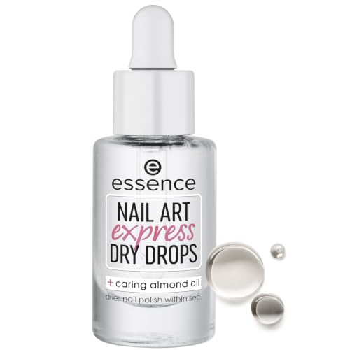
Essence
Nail Art Express Dry Drops
nail polish · 8 ml · 5 ingredients
- Fragrance-free
- No essential oils
- With vitamin e
When to see a professional
Common scattered white spots require no medical attention and will resolve spontaneously. See a GP or dermatologist if: full-width white bands appear across multiple nails (possible Mees' lines); multiple nails show a consistent white pattern suggesting systemic disease; you have symptoms of liver, kidney, or heart disease; white surface changes are present that scrape off (possible superficial onychomycosis); or you have been exposed to heavy metals. Heavy metal poisoning is a medical emergency — seek urgent assessment if arsenic or thallium exposure is possible.
Frequently asked questions
Are white spots on nails really caused by calcium deficiency?
No. The calcium deficiency explanation is a persistent myth with no clinical evidence to support it. White spots (leukonychia punctata) are caused by minor trauma to the nail matrix — the growth zone beneath the cuticle — from nail biting, cuticle manipulation, or everyday knocks. The spots appear weeks after the injury because the damaged cells travel with the growing nail plate. No published study has demonstrated a causal link between dietary calcium or zinc deficiency and leukonychia punctata. The spots resolve without supplementation as the nail grows.
When are white spots on nails a sign of something serious?
Common scattered white spots (leukonychia punctata) are benign. White changes that warrant evaluation include: full-width transverse white bands (Mees' lines) — associated with arsenic or thallium poisoning, chemotherapy, or severe systemic illness; paired horizontal bands on multiple nails (Muehrcke's lines) — a sign of low blood albumin in liver, kidney, or heart disease; a proximal white zone with a narrow pink distal band (Terry's nails) — associated with liver cirrhosis, heart failure, or chronic kidney disease; and white marks on multiple nails simultaneously without any history of trauma. These patterns require clinical evaluation and blood tests.
What causes Mees' lines (white bands that cross the whole nail)?
Mees' lines are transverse white bands running fully across the nail plate. They are a recognized sign of arsenic or thallium poisoning, as well as systemic events that temporarily disturb nail matrix function — including some chemotherapy drugs, renal failure, Hodgkin's lymphoma, and severe acute infections. The lines are effectively a timeline: because the nail grows at a known rate, the distance of a Mees' line from the cuticle indicates roughly when the systemic event occurred. Anyone with Mees' lines in the absence of an obvious cause (such as chemotherapy) should be evaluated urgently by a clinician.
How long do white spots on nails take to go away?
Leukonychia punctata (small white spots from trauma) will travel forward with the nail plate at the rate of approximately 3 mm per month for fingernails, and 1–1.5 mm per month for toenails. A white spot that appears near the mid-nail will typically reach the free edge and be clipped away in 2–3 months; one that appears near the cuticle may take 4–6 months. No treatment speeds up the process. Continued nail biting or matrix trauma will create new spots before old ones grow out.
Can superficial white onychomycosis be mistaken for ordinary white spots?
Yes — superficial white onychomycosis (SWO) produces chalky white patches on the outer surface of the nail plate that can be confused with leukonychia punctata. The key distinguishing feature is that the white material in SWO can be scraped off the nail surface with a file or curette, whereas true leukonychia punctata is inside the nail plate and cannot be scraped away. SWO is a fungal infection (often caused by Trichophyton mentagrophytes on toenails) and responds to topical or oral antifungal treatment — unlike leukonychia punctata, which requires no treatment.
Health information, not medical advice.
Beauty Comparison is an independent editorial publication. This page is researched and sourced to the authorities listed below, but it is general information — not a diagnosis or a substitute for personalised advice from a qualified doctor, dermatologist or pharmacist. If your symptoms are severe, spreading, painful, or not improving, see a healthcare professional.Sources
- 01White Nail — Leukonychia: An Overview — DermNet — DermNet NZ, 2023-01-01. Classification of leukonychia into true, apparent, and pseudo forms; causes of leukonychia punctata (nail matrix trauma), Mees' lines (heavy metal poisoning, systemic illness), Muehrcke's lines (hypoalbuminaemia), Terry's nails (liver/cardiac/renal disease), and pseudoleukonychia (fungal infection, cosmetics).
- 02Leukonychia: What Can White Nails Tell Us? — PMC / NIH, 2022-01-01. Comprehensive review of leukonychia classification; leukonychia punctata caused by abnormal matrix keratinization from trauma; Mees' lines associated with arsenic, thallium, chemotherapy, renal failure; mineral deficiency not an established cause of leukonychia punctata; true vs. apparent distinction (pressure-blanching test).
- 03White Spots on Nails (Leukonychia): Causes & Treatment — Cleveland Clinic, 2022-01-01. Leukonychia punctata most commonly caused by nail or cuticle trauma; calcium and mineral deficiency not confirmed causes; Terry's nails, Muehrcke's lines, and Lindsay's nails as apparent leukonychia patterns associated with systemic disease.
- 04Nail as a Window of Systemic Diseases — PMC / NIH, 2015-01-01. Terry's nails associated with liver cirrhosis, chronic heart failure, type 2 diabetes, chronic kidney disease; Muehrcke's lines linked to hypoalbuminaemia (serum albumin below 2.2 g/dL) and nephrotic syndrome; half-and-half nails (Lindsay's nails) seen in chronic renal failure.