Seborrheic Dermatitis on the Face: Causes & Treatment
The short answer
Facial seborrheic dermatitis is a chronic inflammatory skin condition driven by Malassezia yeast and excess.
What Is Seborrheic Dermatitis on the Face?
Seborrheic dermatitis (SD) is a common, chronic, relapsing inflammatory skin condition affecting sebaceous gland-rich areas of the body — and the face is one of its most visible and troublesome sites. On the face, it produces patches of red or discolored, greasy-looking skin covered with yellowish or white flakes. The nasolabial folds (the creases running from the nose to the corners of the mouth), eyebrows, eyelid margins, and the glabella (the area between the brows) are the characteristic facial zones.
The condition sits on a spectrum with dandruff: dandruff is the uninflamed scalp variant, while seborrheic dermatitis involves visible inflammation. Both share the same underlying biology.
Seborrheic dermatitis affects an estimated 3–12% of the general population, with a clear predominance in males. It follows a biphasic age pattern — infants under three months (where it presents as "cradle cap") and adults from late adolescence onward, peaking again after age 50. For individuals with HIV or AIDS, prevalence climbs dramatically to 30–83%, sometimes appearing as widespread, treatment-resistant disease that can serve as an AIDS-defining clinical marker.
Why the Face? The Biology of Seborrheic Dermatitis
The Malassezia–Sebum Axis
Seborrheic dermatitis does not have a single cause, but the central mechanism is well established. The face — along with the scalp and upper chest — is rich in sebaceous glands. Malassezia yeast (principally M. globosa and M. restricta) lives on everyone's skin as a normal commensal, but in susceptible individuals its overgrowth triggers a pathological cascade:
- Lipase and phospholipase production. Malassezia secretes enzymes that hydrolyze sebum triglycerides, releasing unsaturated free fatty acids — particularly oleic and arachidonic acid.
- Barrier disruption. These fatty acids cause aberrant keratinocyte differentiation and disrupt the stratum corneum, creating a compromised skin barrier.
- Inflammatory cascade. Damaged keratinocytes release pro-inflammatory cytokines (IL-1α, IL-6, IL-8, TNF-α), driving the redness, scaling, and itch that define the condition.
Critically, it is not Malassezia colonisation per se that causes seborrheic dermatitis — the yeast is present on virtually all adults. Rather, it is an abnormal immune response to Malassezia metabolites in a subset of individuals whose skin barrier and inflammatory regulation are susceptible.
Why Some People and Not Others?
No single genetic marker is established, but several factors increase susceptibility:
- Neurological conditions — Parkinson's disease, epilepsy, spinal cord injury, Down syndrome, and stroke recovery are all associated with increased SD prevalence, likely because abnormal neurological signaling affects sebaceous gland regulation and immune tone.
- Immunosuppression — HIV infection, organ transplant recipients, and lymphoma dramatically increase risk and severity.
- Mood disorders — Depression, bipolar disorder, and anorexia nervosa are independently associated with SD, possibly via stress-axis effects on immunity and sebum production.
- Medications — Several oral drugs are associated with flares, including lithium, haloperidol, fluorouracil, griseofulvin, and psoralen.
- Environmental triggers — Cold, dry weather reliably worsens SD; warm humid weather often produces remission. UV light exposure can have a paradoxical mild anti-inflammatory benefit.
- Heavy alcohol use — Associated with increased SD risk via effects on immune function.
What Facial Seborrheic Dermatitis Looks Like
Classic Presentation
The signature appearance is erythematous (red or pink) patches with greasy, yellowish-white scales on a background of oily skin. The lesions are typically bilateral and symmetrical — appearing on both sides of the nose simultaneously, or on both eyebrows — and have ill-defined borders.
Key facial locations:
| Location | Typical Appearance |
|---|---|
| Nasolabial folds | Salmon-pink, greasy, scaly patches in the skin creases |
| Eyebrows and glabella | Yellowish scaling on and between the brows; may extend to the forehead as "corona seborrheica" |
| Eyelid margins (blepharitis) | Yellow crusting between eyelashes; eyelids appear scaly, swollen, and red |
| Ears and retroauricular skin | Scaling inside the ear canal and in the crease behind the ear |
| Beard and mustache area | Greasy, flaking skin beneath facial hair |
| Forehead and hairline | Scaly patches at the scalp–forehead junction |
Symptoms vary from mild flaking with minor itch to intense burning and itch with thick, adherent scale. The condition frequently affects the face and scalp simultaneously.
Presentation on Darker Skin Tones
Seborrheic dermatitis looks different — and is more often misdiagnosed — in people with darker skin tones. The AAD notes that in this group, the rash may:
- Present as hypopigmented (lighter than the surrounding skin) rather than red patches
- Produce petal-shaped or annular (ring-like) scaly lesions, particularly in Black patients
- Leave post-inflammatory hyperpigmentation (dark marks) that can persist long after active disease is controlled
The absence of visible redness should not be taken as a sign that seborrheic dermatitis is absent.
How It Differs from Similar Conditions
Facial seborrheic dermatitis is frequently confused with other conditions — the differential diagnosis matters because treatment differs:
| Condition | Key Distinguishing Features |
|---|---|
| Rosacea | Central facial redness with visible blood vessels; no greasy scale; triggered by heat, alcohol, spicy food |
| Facial psoriasis | Thick, silvery (not greasy) scale; sharper borders; often has associated nail or joint changes |
| Atopic dermatitis (eczema) | Usually itchier; tends to follow skin creases rather than sebaceous zones; often with personal/family atopy history |
| Contact dermatitis | Clear exposure history; may not respect sebaceous distribution; patch testing useful |
| Tinea faciei | Asymmetrical; sharper-edged ring-shaped lesion; confirmed by fungal scraping/culture |
| Rosacea + seborrheic dermatitis | Can co-exist — approximately 20–25% of rosacea patients also have SD |
Treatment: The Evidence
There is no cure for seborrheic dermatitis. The goal of treatment is to control active disease and then maintain remission with ongoing, often lifelong, management. Treatment is matched to severity.
First-Line: Topical Antifungals
Antifungal agents that reduce Malassezia load are the cornerstone of facial SD treatment.
Ketoconazole 2% cream is the best-evidenced agent. Seven double-blind, randomized, vehicle-controlled trials have evaluated ketoconazole for SD; the largest trial (N=575) showed excellent clinical results in 88% of patients. Applied twice daily for active disease; once or twice weekly for maintenance. Ketoconazole 1% and 2% foam (Extina, Xolegel) are FDA-approved alternatives.
Ciclopirox 1% cream is supported by five randomized controlled trials and has comparable efficacy and tolerability to ketoconazole. Applied twice daily for 4+ weeks. It works via a different mechanism (chelation of polyvalent metal cations essential to fungal enzyme function) and is a useful alternative if ketoconazole is not tolerated.
Other topical antifungals with supporting evidence:
- Miconazole cream (1–2× daily)
- Clotrimazole cream
- Terbinafine cream (limited but positive evidence for face and body)
Topical Corticosteroids: Short-Burst Use Only
Mild-to-moderate corticosteroids (hydrocortisone 1%, desonide 0.05%) are effective for rapid symptom relief — particularly itch and acute flares. However, long-term use on the face is contraindicated because of well-documented risks:
- Skin atrophy (thinning)
- Steroid-induced rosacea
- Periorificial dermatitis
- Telangiectasia (visible blood vessels)
- Rebound flares upon discontinuation
Current evidence-based practice: combine a mild corticosteroid for 1–2 weeks at flare initiation with a topical antifungal that is then continued for maintenance; the corticosteroid is discontinued, not the antifungal.
Calcineurin Inhibitors: The Steroid-Free Anti-Inflammatory
Topical calcineurin inhibitors (TCIs) — pimecrolimus 1% cream (Elidel) and tacrolimus 0.1% ointment (Protopic) — provide anti-inflammatory action without steroid-associated risks, making them suitable for the face and eyelid margins where chronic steroid use is particularly problematic.
Evidence: a double-blind, randomized, vehicle-controlled trial (N=96) of pimecrolimus 1% demonstrated superiority to vehicle within 2 weeks. Multiple open-label studies support facial application.
Note: both agents carry an FDA black box warning regarding a theoretical risk of lymphoma or skin cancer based on animal data; no causal relationship has been established in humans, and major dermatology guidelines consider the risk acceptable for the indicated uses. Discuss with your dermatologist.
Roflumilast 0.3% Foam (Zoryve): The Newest Option
Roflumilast foam 0.3%, a phosphodiesterase-4 (PDE4) inhibitor, received FDA approval in 2024 for the topical treatment of seborrheic dermatitis in patients aged 9 years and older. It works by inhibiting PDE4, reducing pro-inflammatory cytokine production without steroids or antifungal activity.
Phase 3 STRATUM trial results (N=457, double-blind, 8 weeks):
- 79.5% of roflumilast patients achieved IGA score 0 (clear) or 1 (almost clear) vs. 58.0% in the vehicle group (p < 0.001)
- Well-tolerated: adverse events occurred in <3.3% of patients, most mild or moderate
- The ceramide-based vehicle (no propylene glycol) minimizes irritation
Roflumilast is an important advance for patients who cannot use steroids or who need a safe, steroid-free maintenance option for the face.
Other Topical Options
| Agent | Mechanism | Evidence | Notes |
|---|---|---|---|
| Lithium gluconate 8% ointment | Modulates immune response | RCT evidence | Twice daily for 8 weeks; particularly studied in Europe |
| Metronidazole 0.75% gel | Anti-inflammatory, antimicrobial | Limited RCT evidence | Off-label; may help milder cases |
| Propylene glycol 20% solution | Reduces Malassezia, loosens scale | Moderate evidence | Useful for acute scale control |
| Zinc pyrithione cream | Antifungal, antimicrobial | Supportive evidence | OTC availability; gentle enough for daily use |
Systemic Treatment: Reserved for Severe or Resistant Disease
For seborrheic dermatitis that fails topical treatment or is unusually widespread, systemic antifungals may be required:
- Oral itraconazole (200 mg daily for 1 week, then 200 mg on 2 consecutive days per month for maintenance): effective for refractory cases
- Oral terbinafine: alternative systemic option with published supporting evidence
Both require monitoring for potential drug interactions and liver effects; systemic treatment is initiated and supervised by a dermatologist.
UVB phototherapy (3 sessions/week, up to 8 weeks) is an option for widespread SD unresponsive to topicals, delivered in a medical setting.
How to Manage Seborrheic Dermatitis on the Face: A Practical Framework
Daily Skin Care
- Use a gentle, fragrance-free, low-pH cleanser morning and evening. Aggressive cleansing strips the already-compromised skin barrier and worsens inflammation.
- Avoid alcohol-based toners and astringents — these trigger flares.
- Moisturize daily with a non-comedogenic, fragrance-free moisturizer. A compromised barrier drives the Malassezia inflammatory cycle; keeping the barrier intact helps limit flares.
- Avoid harsh exfoliants during active disease — physical scrubs and potent acids worsen inflammation.
Triggers to Minimize
While individual triggers vary, the most consistent include:
- Cold, dry weather (use a humidifier in winter)
- Psychological stress — though stress does not cause SD, it reliably worsens active disease
- Alcohol consumption
- Harsh or heavily fragranced skincare products
- Extended fatigue and sleep deprivation (the AAD specifically cites lack of sleep as a trigger in severe adult SD)
- Prolonged UV exposure (some beneficial short-term effect, but sunburn-level exposure worsens the barrier)
Maintenance Strategy
Because seborrheic dermatitis is a chronic, relapsing condition, the goal after achieving control is to prevent relapse, not to stop all treatment. Standard maintenance approaches:
- Intermittent antifungal use (ketoconazole or ciclopirox cream 1–2× per week) even when the skin appears clear
- Roflumilast foam as an ongoing steroid-free maintenance option
- Calcineurin inhibitors on flare days to dampen early inflammation before it escalates
Seborrheic Dermatitis in Special Populations
People with HIV or Immunosuppression
SD in HIV-positive individuals tends to be more severe, more treatment-resistant, and more widespread, and may not respond to standard topical therapy alone. Systemic antifungals are more frequently required. SD severity can mirror immune status, and sudden severe flares in a previously controlled patient warrant an underlying assessment.
People with Parkinson's Disease
There is a particularly well-documented association between Parkinson's disease and seborrheic dermatitis — multiple studies confirm higher prevalence and greater severity in this population. The mechanism likely involves autonomic dysfunction affecting sebaceous gland output. Management is the same as in the general population, but maintenance therapy is usually lifelong.
Eyelid Involvement (Blepharitis)
When SD involves the eyelid margins, blepharitis results — scaling, crusting, and inflammation at the lid edges that can affect vision comfort. Management requires:
- Warm compresses applied for 5–10 minutes to loosen crusts
- Gentle cleaning of the lash line with diluted baby shampoo or a dedicated lid-hygiene product
- Physician-supervised application of ketoconazole or ciclopirox to the lid margins (avoiding direct eye contact)
- Ophthalmology referral if symptoms affect vision, the cornea, or are severe
Common Myths About Facial Seborrheic Dermatitis
"It's caused by not washing my face properly." Seborrheic dermatitis is not caused by poor hygiene. The Malassezia yeast responsible for it lives on virtually every human's skin. In fact, over-washing with harsh cleansers disrupts the skin barrier, worsening the condition. Twice-daily gentle cleansing is both sufficient and appropriate.
"I can eat my way out of it." There is no strong clinical evidence that dietary changes reliably treat or prevent seborrheic dermatitis. Some individuals report improvement after reducing sugar or dairy, but these observations are anecdotal; no randomized controlled trial has demonstrated diet as an effective primary treatment for SD. Treating the skin barrier and Malassezia directly is the evidence-based approach.
"Seborrheic dermatitis means I have HIV." Seborrheic dermatitis is common in the general population (3–12%) and affects the vast majority of people who have no immunodeficiency whatsoever. Severe, sudden-onset, treatment-resistant SD in an adult with risk factors may warrant an HIV test as part of a workup — but SD in isolation is neither a specific nor sensitive indicator of HIV status.
"I just need a stronger steroid to clear it." Potent topical steroids used long-term on the face cause irreversible skin thinning, steroid rosacea, and telangiectasia. The solution is not strength but duration: a mild corticosteroid for a brief initial burst combined with antifungal maintenance is far safer and more effective long-term than escalating steroid potency.
Products that fit: sensitive skin
Picked by code from each product's full ingredient list.

Anua
3 Ceramide Panthenol Moisture Barrier Cream
moisturizer · 100 ml · 63 ingredients
- Fragrance-free
- No essential oils
- No drying alcohol
- No MI/MCI
- No menthol, camphor and witch hazel

Naturium
Marshmallow Root Barrier Balm
moisturizer · 50 g · 58 ingredients
- Fragrance-free
- No essential oils
- No drying alcohol
- No MI/MCI
- No menthol, camphor and witch hazel
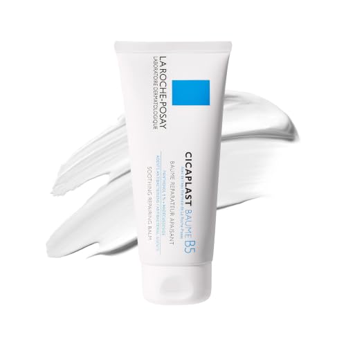
La Roche-Posay
Cicaplast Baume B5 Soothing Repair
moisturizer · 100 ml · 33 ingredients
- Fragrance-free
- No essential oils
- No drying alcohol
- No MI/MCI
- No menthol, camphor and witch hazel

Beauty of Joseon
Red Bean Water Gel
moisturizer · 99.9 ml · 58 ingredients
- Fragrance-free
- No essential oils
- No drying alcohol
- No MI/MCI
- No menthol, camphor and witch hazel
When to see a professional
See a dermatologist if seborrheic dermatitis does not improve after 4 weeks of consistent OTC antifungal use; if it involves the eyelids or is affecting vision comfort; if the skin becomes secondarily infected (increasing warmth, pus, crusting beyond scaling); if the rash is unusually severe, widespread, or appeared suddenly in an adult with no prior history — this warrants an assessment for underlying immunosuppression including HIV; if the condition is significantly affecting quality of life or mental health; or if you are unable to distinguish the rash from psoriasis, rosacea, or another condition that requires a different treatment approach. Prescription options (stronger antifungals, calcineurin inhibitors, roflumilast) substantially outperform OTC-only management for many patients.
Frequently asked questions
What does the latest evidence say about treating seborrheic dermatitis on the face in 2026?
The evidence base for facial seborrheic dermatitis has expanded meaningfully with roflumilast 0.3% foam (Zoryve), which received FDA approval in 2024 as a steroid-free PDE4 inhibitor. In the phase 3 STRATUM trial (N=457), 79.5% of patients using roflumilast achieved clear or near-clear skin at 8 weeks compared to 58% with vehicle — a statistically significant and clinically meaningful result. The established standard — ketoconazole 2% cream (backed by seven double-blind RCTs) and ciclopirox 1% cream — remains first-line, with calcineurin inhibitors (pimecrolimus, tacrolimus) as effective steroid-sparing anti-inflammatories. Short-burst mild corticosteroids are still used for acute control, but the focus has shifted toward nonsteroidal long-term maintenance to avoid cumulative steroid harms on facial skin.
Why does seborrheic dermatitis appear specifically on the face and scalp rather than the arms or legs?
The distribution of seborrheic dermatitis directly follows the density of sebaceous glands. The scalp, mid-face (nasolabial folds, eyebrows, glabella), ear canal, and upper chest have the highest sebaceous gland density in the body — and Malassezia yeast that drives SD feeds exclusively on sebum-derived lipids. Body sites with low sebaceous activity (arms, legs, palms, soles) rarely develop seborrheic dermatitis.
Is facial seborrheic dermatitis the same as dandruff?
They share the same underlying biology — Malassezia yeast metabolizing sebum — but dandruff is the uninflamed scalp variant (flaking without significant redness or itch), while seborrheic dermatitis involves visible inflammation, scaling, and often itch on the scalp, face, or body. Many people with facial seborrheic dermatitis also have concurrent dandruff. Treatment strategies overlap: ketoconazole and ciclopirox treat both, though delivery forms differ (cream for the face, shampoo for the scalp).
Can seborrheic dermatitis on the face be permanently cured?
No. Seborrheic dermatitis is a chronic, relapsing condition in adults — the predisposition is lifelong. The goal is sustained remission through maintenance therapy, not a one-time cure. Many patients achieve long, symptom-free periods with consistent intermittent antifungal use (e.g., ketoconazole cream 1–2× per week even between flares). Infants with cradle cap are the exception: the condition typically resolves within weeks to months as the influence of maternal hormones on sebaceous glands wanes.
Is seborrheic dermatitis contagious?
No. Seborrheic dermatitis is not contagious. The Malassezia yeast involved is a normal part of every human's skin microbiome. The condition arises from an individual's own immune response to Malassezia metabolites in the context of high sebum production — it cannot be passed from person to person through contact.
Can seborrheic dermatitis cause permanent skin damage or scarring?
Active seborrheic dermatitis itself does not typically cause scarring. However, prolonged uncontrolled disease can result in post-inflammatory hyperpigmentation (dark marks), particularly in people with darker skin tones, and these marks may take months to fade. Additionally, prolonged inappropriate use of potent topical steroids on the face to manage SD can cause permanent skin thinning and visible blood vessels — which is a treatment complication, not a disease complication. Prompt, appropriate management prevents both.
Health information, not medical advice.
Beauty Comparison is an independent editorial publication. This page is researched and sourced to the authorities listed below, but it is general information — not a diagnosis or a substitute for personalised advice from a qualified doctor, dermatologist or pharmacist. If your symptoms are severe, spreading, painful, or not improving, see a healthcare professional.Sources
- 01Seborrheic Dermatitis: Overview, Causes, Symptoms, and Treatment — American Academy of Dermatology (AAD). Prevalence across ages and races; biphasic peak (infancy and adulthood 50+); male predominance; association with HIV, Parkinson's, psoriasis, rosacea; chronic relapsing course; no permanent cure.
- 02Seborrheic Dermatitis: Causes and Risk Factors — American Academy of Dermatology (AAD), 2024-01-01. Malassezia yeast and sebum as the two central factors; association with neurological disorders, HIV, lymphoma, mood disorders, Down syndrome, stroke, organ transplant, heavy alcohol use; medication triggers (lithium, haloperidol, fluorouracil, griseofulvin, psoralen); cold/dry weather as seasonal trigger; lack of sleep and stress as flare factors.
- 03Seborrheic Dermatitis: Diagnosis and Treatment — American Academy of Dermatology (AAD), 2024-01-01. Topical antifungals (ketoconazole), corticosteroids, calcineurin inhibitors (Elidel, Protopic), ciclopirox shampoo, propylene glycol solution, roflumilast foam (Zoryve, 68.3% clear/almost clear at 8 weeks), oral itraconazole/terbinafine for severe cases, UVB phototherapy.
- 04Seborrhoeic Dermatitis: Causes and Treatment — DermNet — DermNet NZ, 2023-06-01. Prevalence 3–12%; Malassezia pathophysiology via lipase/phospholipase production; facial distribution (nasolabial folds, eyebrows, eyelid margins, ears); blepharitis; differential diagnosis from psoriasis, rosacea, atopic dermatitis, tinea, contact dermatitis; ketoconazole and ciclopirox as first-line; mild corticosteroids for short-burst use; calcineurin inhibitors for steroid-sparing; roflumilast 0.3% foam FDA-approved; systemic options for resistant disease; darker-skin presentation as hypopigmented petal-shaped lesions.
- 05An Overview of the Diagnosis and Management of Seborrheic Dermatitis — PMC / NCBI (Cureus), 2022-08-01. Multifactorial pathogenesis: Malassezia spp., sebaceous gland activity, immunosuppression, neurogenic and iatrogenic factors; HIV prevalence 30–83%; Level A evidence for ketoconazole 1–2%, ciclopirox 1%, mild corticosteroids, lithium succinate/gluconate; calcineurin inhibitors off-label Level A/B; management targeting sebum modulation, Malassezia reduction, and inflammation control.
- 06Seborrheic Dermatitis and Dandruff: A Comprehensive Review — PMC / NCBI (Journal of Clinical & Investigative Dermatology), 2015-12-01. Affects 1–3% of general adult population; M. globosa and M. restricta are primary species; pathophysiology: lipase hydrolyzes sebum → unsaturated fatty acids → aberrant keratinocyte differentiation → IL-1α, IL-6, IL-8, TNF-α cytokine release; facial sites: nasolabial folds, upper lip, eyelids, eyebrows, corona seborrheica; ketoconazole 2% cream with 88% excellent results in largest trial (N=575); evidence table for topical treatments including ketoconazole, ciclopirox, hydrocortisone, desonide, pimecrolimus, tacrolimus, lithium gluconate.
- 07Adult Seborrheic Dermatitis: A Status Report on Practical Topical Management — PMC / NCBI (Journal of Clinical & Aesthetic Dermatology), 2011-05-01. Ketoconazole: 7 double-blind RCTs; 88% excellent results in N=575 trial; ketoconazole 2% superior to 1% and to selenium sulfide 2.5%; ciclopirox: 5 double-blind RCTs; pimecrolimus 1%: double-blind RCT (N=96) showing superiority to vehicle by week 2; evidence-based recommendation: short-course corticosteroid combined with nonsteroidal agent for moderate-to-severe flare; intermittent antifungal for maintenance; combination outperforms monotherapy.
- 08Roflumilast Foam (Zoryve) for Seborrheic Dermatitis — FDA Approval 2024 — PubMed / NCBI, 2024-03-01. Roflumilast 0.3% foam FDA-approved 2024 for seborrheic dermatitis in patients ≥9 years; STRATUM phase 3 trial (N=457, 8 weeks): 79.5% of roflumilast patients achieved IGA success (clear/almost clear) vs. 58.0% vehicle (p<0.001); adverse events <3.3%, mild-to-moderate; ceramide-based vehicle minimizes irritation.