Scalp Psoriasis vs. Seborrheic Dermatitis: Key Differences
The short answer
Scalp psoriasis has thick silvery plaques on clearly red skin; seborrheic dermatitis has yellowish.
Why These Two Conditions Are So Often Confused
Scalp psoriasis and seborrheic dermatitis share enough surface features — both cause an inflamed, scaly, itchy scalp — that even dermatologists find them challenging to distinguish in some cases. They are the most common causes of chronic scalp scaling in adults, and they can coexist. Understanding the distinctions, and knowing when to seek expert assessment, is clinically important because the treatments are different.
The Fundamentally Different Causes
Scalp Psoriasis: An Autoimmune Process
Scalp psoriasis is an immune-mediated inflammatory disorder. In genetically predisposed individuals, dendritic cells in the skin release IL-23, which drives the maturation of Th17 cells that then produce IL-17A — the principal cytokine responsible for the hyperproliferation of keratinocytes seen in psoriatic plaques. Skin cells that normally complete their 28–30 day maturation cycle in psoriasis do so in just 3–4 days, producing the thick scale characteristic of the condition. The underlying cause is intrinsic immune dysregulation, not a microorganism.
Seborrheic Dermatitis: A Yeast-Triggered Inflammatory Response
Seborrheic dermatitis affects 3–10% of the general population and is driven by the commensal yeast Malassezia — particularly M. globosa and M. restricta. These yeasts are present on the scalps of virtually all adults; the inflammatory response in seborrheic dermatitis is a heightened, aberrant reaction to Malassezia metabolites (primarily free fatty acids generated when the yeast's enzymes break down scalp sebum). The result is scalp inflammation that is moderate in severity and differs structurally from the psoriatic autoimmune cascade. Antifungal treatment that suppresses Malassezia effectively controls seborrheic dermatitis.
This fundamental difference in cause explains why treatments targeting Malassezia (antifungals) clear seborrheic dermatitis but are ineffective in psoriasis.
Clinical Comparison: The Key Distinguishing Features
| Feature | Scalp Psoriasis | Seborrheic Dermatitis |
|---|---|---|
| Cause | Autoimmune (IL-23/IL-17) | Malassezia yeast-triggered inflammation |
| Scale appearance | Thick, dry, silvery-white | Yellowish, greasy, softer |
| Plaque definition | Well-demarcated, sharp borders | Diffuse, poorly defined, blends into normal scalp |
| Underlying skin color | Prominently red, raised | Pale pink to mild erythema |
| Scalp coverage pattern | Focal plaques or diffuse; can be patchy | Diffuse involvement of the scalp |
| Extension beyond hairline | Common — forehead, nape, ears | Possible at hairline but typically less prominent |
| Face involvement | Face not typical (forehead extension only) | Common — eyebrows, nasolabial folds, beard, chest |
| Nail changes | Common (pitting, onycholysis) | Absent |
| Joint involvement | Psoriatic arthritis in ~30% of psoriasis patients | Not associated with joint disease |
| Body site lesions | Can coexist with elbows, knees, lower back plaques | Typically confined to sebaceous regions |
| Itch | Often moderate to severe | Mild to moderate |
| Burning / soreness | Common | Less typical |
| Response to antifungals | No clinically meaningful response | Good response to ketoconazole, ciclopirox |
| Response to topical corticosteroids | Good, but short-term in isolation | Good for acute control; not for long-term maintenance |
The Four Clinical Clues to Focus On
1. Scale color and greasiness
This is the most reliably distinguishing visual feature:
- Psoriasis scale: thick, dry, silvery-white — often compared to candle wax or mica
- Seborrheic dermatitis scale: thinner, softer, yellowish or honey-colored, with a slightly greasy or waxy texture reflecting the sebum-rich environment in which Malassezia thrives
2. Plaque definition
Part the hair and examine the edges of the inflamed area:
- Psoriasis: the border between inflamed and normal scalp skin is sharp and well-defined — you can clearly see where the plaque ends
- Seborrheic dermatitis: the transition from inflamed to normal skin is gradual and indistinct; the scaling blends into the surrounding scalp without a clean margin
3. Face and body involvement
- Psoriasis: may extend onto the forehead (at the hairline), behind the ears, and the nape of the neck; body site plaques on elbows, knees, lower back, and nails are common
- Seborrheic dermatitis: frequently affects the face in characteristic sebaceous areas — the eyebrows, the sides of the nose, nasolabial folds, beard area — and the chest; nail involvement is not associated
4. Response to antifungal treatment
This is diagnostically useful if there is uncertainty:
- Using a ketoconazole shampoo twice weekly for 4 weeks and observing the response can be informative: good clearance supports seborrheic dermatitis; no response (or worsening) increases suspicion for psoriasis
Sebopsoriasis: When the Two Overlap
Sebopsoriasis (also called seborrhoeic psoriasis) is a recognized clinical entity in which features of both conditions coexist in the same patient. It typically presents with:
- Scale that is somewhat silvery-white but with a yellowish or greasy component
- Plaques that are less sharply defined than typical psoriasis
- Distribution across the scalp and face, mimicking seborrheic dermatitis
- Possible coexistence of typical psoriatic plaques elsewhere on the body
The overlap is thought to reflect either genuinely concurrent conditions or a disease phenotype that sits between the two diagnoses. Histopathology may be needed to clarify.
Confirming the Diagnosis: Dermoscopy and Biopsy
When clinical features are ambiguous, two tools are used:
Trichoscopy (dermoscopy of the scalp)
Trichoscopy reveals distinctive vascular and scale patterns:
- Scalp psoriasis: red dots (dilated capillary loops), hairpin vessels, and red globular rings — reflecting the papillary dermal vascular changes of psoriatic plaques
- Seborrheic dermatitis: arborizing red lines and comma vessels — a different vascular pattern reflecting the more diffuse, Malassezia-associated inflammation
Scalp biopsy (histopathology)
When trichoscopy and clinical features are insufficient, biopsy is definitive. In a study comparing histopathological features (PMC4969470):
Features significantly more common in psoriasis:
- Mounds of parakeratosis with neutrophils (100% of psoriasis cases)
- Spongiform micropustules of Kogoj (33.3% vs 0% in seborrheic dermatitis)
- Clubbed and evenly elongated rete ridges (100% of psoriasis cases)
Features significantly more common in seborrheic dermatitis:
- Follicular plugging (70% vs 26.7% in psoriasis)
- Shoulder parakeratosis (40% vs 6.7%)
- Prominent lymphocytic exocytosis (45% vs 6.7%)
- Marked spongiosis (21.1% vs 0% in psoriasis)
Treatment: How the Approaches Differ
For Scalp Psoriasis
First-line treatment targets the autoimmune inflammatory cascade:
- Topical corticosteroids (shampoos, foams, gels, solutions) — most commonly prescribed; clobetasol propionate for stubborn cases
- Topical vitamin D analogues (calcipotriol) — normalize keratinocyte proliferation; most effective combined with a corticosteroid
- Salicylic acid — soften thick scale to improve penetration of other treatments
- Systemic or biologic therapy for moderate-to-severe disease (see Scalp Psoriasis Guide)
For Seborrheic Dermatitis
Treatment targets Malassezia and associated inflammation:
- Ketoconazole shampoo (1%–2%) — most evidence-backed antifungal; used twice weekly for control, then once weekly for maintenance
- Ciclopirox shampoo — antifungal with additional anti-inflammatory activity; effective maintenance option
- Selenium sulfide shampoo — reduces Malassezia
- Zinc pyrithione shampoo — antibacterial and antifungal; widely available OTC
- Topical corticosteroids — useful for acute flares but not appropriate for long-term maintenance in seborrheic dermatitis given the chronic, relapsing nature of the condition
- The condition tends to relapse after treatment is stopped; maintenance with antifungal shampoo is typically necessary
When to See a Dermatologist
- Medicated shampoos (whether antifungal or OTC coal tar/salicylic acid) have not produced meaningful improvement after 6 weeks of consistent use
- There is visible redness with sharply defined plaque borders — escalation to prescription psoriasis treatment is needed
- The scaling extends beyond the hairline onto the forehead, behind the ears, or nape
- Facial involvement is present (eyebrows, nasolabial folds)
- Joint pain or stiffness is occurring alongside scalp scaling — possible psoriatic arthritis warrants evaluation
- Significant hair shedding is occurring
- You are uncertain which condition you have
Products that fit: psoriasis
Picked by code from each product's full ingredient list.

Eucerin
Dermo Capillaire Anti-dandruff Shampoo
shampoo · 250 ml · 15 ingredients
- Fragrance-free
- No drying alcohol
- With urea
- With lactic acid

The Body Shop
Tea Tree Shampoo
shampoo · 400 ml · 22 ingredients
- Fragrance-free
- No drying alcohol
- With salicylic acid
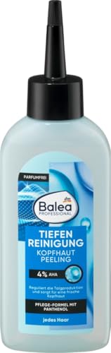
Balea
Professional Scalp Scrub Deep Cleansing
scalp treatment · 150 ml · 10 ingredients
- Fragrance-free
- No drying alcohol
- With glycolic acid
- With panthenol

OGX
ProGrowth + Peptide Scalp Serum
scalp treatment · 10 ml · 16 ingredients
- Fragrance-free
- No drying alcohol
- With niacinamide
- With panthenol
- With peptides
Frequently asked questions
What is the best current evidence for distinguishing scalp psoriasis from seborrheic dermatitis?
Clinically, scale character (silvery-white and dry vs. yellowish-greasy), plaque definition (sharp vs. diffuse), and body site involvement remain the most reliable discriminators. Trichoscopy has added a non-invasive objective tool: psoriasis characteristically shows red dots, hairpin vessels, and red globular rings; seborrheic dermatitis shows arborizing red lines and comma vessels (PMC4318061). Histopathology remains the diagnostic gold standard — mounds of parakeratosis with neutrophils and Kogoj micropustules in psoriasis; follicular plugging and marked spongiosis in seborrheic dermatitis (PMC4969470). A 2022 deep-learning dermoscopy model (PMC9669613) achieved 96.1% sensitivity and 88.2% specificity — comparable to expert dermatologists — but does not replace clinical evaluation.
Can scalp psoriasis and seborrheic dermatitis occur at the same time?
Yes. Sebopsoriasis is a recognized overlap presentation in which features of both conditions coexist, making clear-cut diagnosis difficult even for experienced dermatologists. Patients may have the greasy yellowish scale of seborrheic dermatitis alongside the sharper red plaques of psoriasis, or an intermediate appearance. Management in overlap cases typically addresses both pathways — antifungal shampoo alongside topical corticosteroids — and dermatologist input is strongly advised.
Is seborrheic dermatitis contagious?
No. Although seborrheic dermatitis is driven by Malassezia yeast, this yeast is a normal commensal present on virtually all adult scalps. The condition is a heightened inflammatory response to Malassezia in susceptible individuals, not an infection that is transmitted between people. It cannot be caught from another person.
Why do antifungal shampoos not work for scalp psoriasis?
Scalp psoriasis is caused by autoimmune immune dysregulation — specifically, dysregulated T-cell signalling via the IL-23/IL-17 axis causing keratinocyte hyperproliferation. Antifungal agents reduce Malassezia, which is the driver of seborrheic dermatitis. Since psoriasis is not driven by any fungal organism, antifungals have no effect on the underlying mechanism and produce no clinically meaningful improvement in psoriatic plaques.
How long does seborrheic dermatitis take to respond to treatment?
Most people with seborrheic dermatitis see significant improvement within 2–4 weeks of using an appropriate antifungal shampoo (ketoconazole, ciclopirox) consistently. However, seborrheic dermatitis is a chronic condition that tends to relapse when treatment is stopped; maintenance therapy with once-weekly antifungal shampoo is typically needed to prevent recurrence. Scalp psoriasis tends to require longer and more sustained treatment, particularly for thick, established plaques.
Health information, not medical advice.
Beauty Comparison is an independent editorial publication. This page is researched and sourced to the authorities listed below, but it is general information — not a diagnosis or a substitute for personalised advice from a qualified doctor, dermatologist or pharmacist. If your symptoms are severe, spreading, painful, or not improving, see a healthcare professional.Sources
- 01Histopathological Differential Diagnosis of Psoriasis and Seborrheic Dermatitis of the Scalp — PMC / NIH, 2016-06-01. Kogoj micropustules (33.3% psoriasis vs 0% SD), mounds of parakeratosis with neutrophils (100% vs 50%), clubbed rete ridges (100% psoriasis) favored psoriasis; follicular plugging (70% SD), shoulder parakeratosis (40%), lymphocytic exocytosis (45%), spongiosis (21.1% SD only) favored seborrheic dermatitis — definitive histopathological criteria for differentiation.
- 02Seborrheic Dermatitis of the Scalp — PMC / NIH, 2015-06-01. Seborrheic dermatitis affects 3–10% of population; Malassezia (M. globosa, M. restricta) drives inflammation via free fatty acids from sebaceous triglycerides; ketoconazole most effective; tends to relapse after treatment; distinguishing features from psoriasis include more prominent erythema and hairline involvement in psoriasis.
- 03Dermoscopic Findings in Scalp Psoriasis and Seborrheic Dermatitis; Two New Signs: Signet Ring Vessel and Hidden Hair — PMC / NIH, 2015-01-01. Trichoscopy distinguishes the two conditions: red dotted globules characteristic of psoriasis; arborizing red lines and comma vessels more characteristic of seborrheic dermatitis.
- 04A Deep Learning-Based Approach Toward Differentiating Scalp Psoriasis and Seborrheic Dermatitis from Dermoscopic Images — PMC / NIH, 2022-11-01. Deep learning model achieved 96.1% sensitivity and 88.2% specificity for differentiating scalp psoriasis vs seborrheic dermatitis on dermoscopy — comparable to expert dermatologist performance.
- 05The Immunologic Role of IL-17 in Psoriasis and Psoriatic Arthritis Pathogenesis — PMC / NIH, 2018-11-01. IL-23/IL-17 axis is the central immunological driver of psoriasis; dendritic cells → Th17 cells → IL-17A → keratinocyte hyperproliferation; explains why antifungal treatment is ineffective for psoriasis.
- 06Scalp Psoriasis: A Complete Overview — DermNet NZ. Scalp psoriasis: silvery-white scale on red well-demarcated plaques; sebopsoriasis recognized overlap entity; differential includes seborrheic dermatitis; treatment ladder from topicals to biologics.
- 07Seborrheic Dermatitis: Causes and Treatment — DermNet NZ. Seborrheic dermatitis: yellowish greasy scales; affects scalp, face (eyebrows, nasolabial folds, beard), chest; Malassezia driven; ketoconazole and ciclopirox effective; antifungal maintenance needed to prevent relapse.