Tinea Capitis (Scalp Ringworm): Symptoms, Causes & Treatment
The short answer
Tinea capitis is a fungal infection of the scalp requiring oral antifungal medication.
What Is Tinea Capitis?
Tinea capitis — commonly called scalp ringworm — is a dermatophyte fungal infection that colonises the skin of the scalp and invades the hair shafts and follicles. Despite the name, no worm is involved; the "ring" refers to the characteristic circular, scaly patches the infection can produce.
It is among the most common fungal infections of childhood worldwide, predominantly affecting children between 3 and 14 years of age, though immunocompromised adults can develop it at any age. The condition is contagious and, if untreated or treated inadequately, can cause permanent hair loss.
Causative Organisms
Tinea capitis is caused by fungi called dermatophytes — specifically genera Trichophyton and Microsporum. The dominant species varies by geography:
- Trichophyton tonsurans — the most common cause in the United States and urban areas of Western Europe; anthropophilic (human-to-human transmission), endothrix invasion pattern
- Microsporum canis — predominates in Europe, New Zealand, and parts of Asia; zoophilic (most often transmitted from cats and dogs), ectothrix invasion pattern
- Trichophyton violaceum — common in Africa and among immigrants from the Middle East and Africa
- Trichophyton schoenleinii — rare; causes the chronic scarring form called favus
How Hair Is Invaded
Dermatophytes invade the hair follicle in two patterns:
| Pattern | What Happens | Key Species |
|---|---|---|
| Endothrix | Spores grow inside the hair shaft; hair breaks at scalp level ("black dot") | T. tonsurans, T. violaceum |
| Ectothrix | Spores coat the outside of the hair shaft; hair breaks above scalp | M. canis, M. audouinii |
| Favus | Chronic crust-forming infection; scarring alopecia risk | T. schoenleinii |
How Tinea Capitis Spreads
The infection is contagious, transmitted by:
- Direct contact with an infected person's scalp (most common)
- Shared objects — combs, brushes, hats, pillowcases, towels
- Infected animals — cats and dogs are the main source of M. canis; cattle for T. verrucosum
- Soil (rare; certain geophilic species)
Children attending schools or nurseries in crowded settings are at higher risk. Warm, humid climates increase prevalence. Contrary to common belief, poor hygiene is not a direct cause — the infection can spread in any hygienic environment through close contact.
Symptoms and Clinical Presentations
Tinea capitis presents in several distinct patterns, broadly divided into non-inflammatory and inflammatory forms.
Non-Inflammatory Forms
Gray-patch tinea capitis Circular patches of dull, grayish scaling on the scalp with hair that breaks just above the surface, leaving short stubs. Multiple patches may coalesce. Itch is variable. This pattern is typical of Microsporum species and is the classic "ringworm" appearance.
Black-dot tinea capitis Associated with endothrix infections (T. tonsurans). Hair breaks at the scalp surface, leaving black dots (remnant hair stumps) within the scaly patch. May mimic alopecia areata or seborrheic dermatitis.
Diffuse scaling Widespread fine scale resembling dandruff, sometimes without obvious alopecia. This presentation is frequently misdiagnosed and under-treated. A useful clinical clue: dandruff in a child under 12 should raise suspicion for tinea capitis.
Inflammatory Forms
Kerion A kerion is the most severe presentation — a boggy, raised, painful inflammatory mass studded with pustules and seeping serous or purulent discharge. It represents a vigorous immune response to the fungal infection rather than bacterial superinfection, though secondary bacterial colonisation can occur. Kerion risks permanent scarring alopecia if antifungal treatment is delayed.
Pustular tinea capitis Multiple follicular pustules across a broader scalp area, sometimes accompanied by enlarged lymph nodes at the back of the neck (posterior cervical lymphadenopathy) — a finding strongly suggestive of tinea capitis in a child.
Favus A rare, chronic form caused by T. schoenleinii: yellow, cup-shaped crusts called scutula form around each hair shaft, producing a honeycomb appearance and a characteristic "mousy" odor. Without treatment, extensive scarring alopecia develops.
Systemic Signs
Posterior or occipital lymphadenopathy is common, particularly with inflammatory forms. Low-grade fever may accompany severe kerion.
Diagnosis
Clinical assessment alone is insufficient — diagnosis should be confirmed, especially before committing to weeks of oral antifungal treatment in a child.
KOH Microscopy
Scalp scrapings or plucked hairs are mounted in potassium hydroxide (KOH) solution and examined under a microscope. Hyphae and arthrospores (inside or on the hair shaft depending on species) confirm dermatophyte infection. Results are available within hours and are positive in the vast majority of symptomatic cases.
Fungal Culture
The gold standard for species identification. Hair clippings or a moistened swab across the affected area is plated on Sabouraud's dextrose agar. Results take 2–4 weeks but are needed to direct antifungal selection (griseofulvin vs terbinafine), particularly where treatment has failed.
Wood's Lamp Examination
An ultraviolet light examination: Microsporum species fluoresce bright green; Trichophyton tonsurans (the most common US species) does not fluoresce, limiting this test's utility in North America.
Trichoscopy (Dermoscopy of the Scalp)
A non-invasive tool showing characteristic patterns — comma hairs, corkscrew hairs, barcode hairs, and broken hair stubs — that allow clinical diagnosis before culture results return.
Treatment
Why Topical Antifungals Fail
Topical antifungals (creams, lotions, solutions) cannot penetrate deeply enough into the hair follicle to eradicate the infection. Tinea capitis always requires systemic oral antifungal treatment for a minimum of 4 weeks, and usually 6–8 weeks or longer.
First-Line Oral Antifungals
Griseofulvin The longest-established treatment and the first-line choice for Microsporum infections:
- Children: 20–25 mg/kg/day (microsize) or 10–15 mg/kg/day (ultramicrosize)
- Adults: 500–1,000 mg/day
- Duration: 6–8 weeks (sometimes up to 12 weeks for Microsporum)
- Absorbed better with a fatty meal; take with milk or a full meal
- Availability has been inconsistent in some regions
Terbinafine Preferred for Trichophyton infections (the most common US cause); weight-based dosing in children:
- <20 kg: 62.5 mg/day
- 20–40 kg: 125 mg/day
-
40 kg: 250 mg/day
- Adults: 250 mg/day
- Duration: 4 weeks (for Trichophyton); may need 8+ weeks for Microsporum, where it is less effective
Clinical evidence supports comparable efficacy between the two agents for Trichophyton species, with a 4-week terbinafine course showing similar outcomes to an 8-week griseofulvin course.
Alternative Agents
- Itraconazole (pulse or continuous dosing): effective for both Trichophyton and Microsporum; useful when griseofulvin is unavailable or not tolerated
- Fluconazole: an alternative when first-line agents cannot be used; generally less effective than terbinafine for Trichophyton
Adjunctive Shampoo Therapy
Antifungal shampoos (2% ketoconazole, 1–2.5% selenium sulfide) do not replace systemic therapy but are strongly recommended twice weekly to:
- Reduce fungal spore shedding from the scalp (reduces household contagion)
- Speed resolution of scalp scaling
- Treat household contacts prophylactically
Managing a Kerion
A kerion is an emergency of sorts — prompt antifungal treatment is the priority. Do not incise a kerion (it is not an abscess in the usual sense; draining it spreads infection and increases scarring risk). A short course of oral corticosteroids (prednisolone) may be added alongside the antifungal to reduce inflammation and lower the risk of scarring alopecia; evidence for this is supportive though not definitive.
Contagion and School Attendance
Tinea capitis is contagious but children do not need to be excluded from school once systemic antifungal treatment has started — provided the scalp is covered and antifungal shampoo use has begun. This is the current recommendation of most pediatric dermatology guidelines.
Household contacts should be examined and treated with antifungal shampoo prophylactically, even if asymptomatic, to prevent re-infection.
Shared objects — combs, hats, pillowcases, hair accessories, headphones — should be disinfected or not shared until the infection is cleared.
Prognosis and Potential Complications
| Outcome | Detail |
|---|---|
| Non-inflammatory forms | Excellent with prompt, adequate oral treatment; hair regrows fully |
| Kerion | Good if treated early; risk of permanent scarring alopecia increases with delayed treatment |
| Favus | High risk of permanent alopecia without early sustained therapy |
| Id reaction (dermatophytide) | A distant, non-infectious rash triggered by immune response to the fungus; appears shortly after starting treatment and resolves without stopping antifungals |
Tinea Capitis vs. Other Scalp Conditions
Tinea capitis is frequently misdiagnosed. Key differentiators:
| Feature | Tinea Capitis | Dandruff / Seborrheic Dermatitis | Alopecia Areata | Scalp Psoriasis |
|---|---|---|---|---|
| Age group | Predominantly children | Any age | Any age | Any age |
| Hair loss | Yes — patchy, with broken hairs | None | Yes — smooth bald patches, no scale | Uncommon unless severe |
| Scalp appearance | Scaly patches, black dots, kerion | Diffuse greasy or dry scale | No scale; smooth skin | Thick silvery-white scale |
| Contagious | Yes | No | No | No |
| Lymphadenopathy | Yes (posterior cervical nodes) | No | No | No |
| Diagnosis | KOH / culture confirms | Clinical | Clinical | Clinical / biopsy |
| Treatment | Oral antifungal (mandatory) | Antifungal shampoos | Observation / steroids / JAK inhibitors | Topical steroids / biologics |
See Ringworm vs. Dandruff on the Scalp for a detailed head-to-head comparison.
Products that fit: itchy, sensitive scalp
Picked by code from each product's full ingredient list.
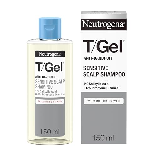
Neutrogena
T/Gel Anti Dandruff Shampoo
shampoo · 150 ml · 15 ingredients
- Fragrance-free
- No essential oils
- No MI/MCI
- With piroctone olamine
- With salicylic acid
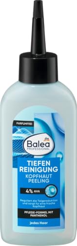
Balea
Professional Scalp Scrub Deep Cleansing
scalp treatment · 150 ml · 10 ingredients
- Fragrance-free
- No essential oils
- No MI/MCI
- With panthenol

OGX
ProGrowth + Peptide Scalp Serum
scalp treatment · 10 ml · 16 ingredients
- Fragrance-free
- No essential oils
- No MI/MCI
- With panthenol

Eucerin
Dermo Capillaire Anti-dandruff Shampoo
shampoo · 250 ml · 15 ingredients
- Fragrance-free
- No essential oils
- No MI/MCI
- With panthenol
When to see a professional
Seek medical attention promptly if a child develops scaly, flaking patches with hair loss or broken hairs on the scalp — tinea capitis cannot be treated with OTC antifungal creams alone and requires a prescription oral antifungal. Urgent assessment is needed for a kerion (painful, boggy, weeping scalp mass) as delayed treatment risks permanent hair loss. See a dermatologist if: the infection fails to improve after completing a full course of prescribed treatment; the diagnosis is uncertain; an immunocompromised person (child or adult) is affected; or a household cluster of cases occurs.
Frequently asked questions
Is tinea capitis treatment still the same in 2026?
Yes — systemic oral antifungal therapy remains the standard of care and the evidence base is unchanged. Griseofulvin and terbinafine are the two first-line agents, with selection guided by the likely causative species. A 2024 pediatric case series and prior head-to-head trials confirm that 4 weeks of terbinafine is comparably effective to 8 weeks of griseofulvin for Trichophyton infections. No newer antifungal has been approved specifically for tinea capitis since these agents, and topical-only treatment is still considered ineffective.
How long does tinea capitis take to clear?
With appropriate systemic treatment, the infection is typically eradicated within 4–8 weeks, though scalp scaling may clear before laboratory confirmation of mycological cure. Terbinafine for Trichophyton infections typically requires 4 weeks. Griseofulvin for Microsporum infections often requires 6–8 weeks, sometimes up to 12. Hair regrowth in non-inflammatory cases begins during treatment and is usually complete within a few months of cure. Kerion requires the same treatment duration but follow-up is essential to assess for scarring.
Can adults get tinea capitis?
Yes, though it is far less common in adults than in children. Adults at higher risk include immunocompromised individuals (HIV, organ transplant recipients, people on long-term corticosteroids), those with close contact with infected children or animals, and people in certain occupational settings. Adult presentations can be atypical and are more frequently missed. The same oral antifungal drugs are used, at adult doses.
Can tinea capitis spread to other family members?
Yes — it is highly contagious. Spread occurs through direct scalp-to-scalp contact and via shared objects (combs, brushes, hats, pillowcases, headphones). Household contacts should be examined and should use an antifungal shampoo (ketoconazole or selenium sulfide) twice weekly prophylactically, even if they have no symptoms. The infected person should not share any hair items until treatment is complete and mycological cure is confirmed.
What is a kerion and how is it different from a regular scalp infection?
A kerion is a severe, boggy, painful inflammatory mass on the scalp with multiple pustules and discharge. It is caused by a vigorous immune response to the dermatophyte infection — not by bacterial superinfection, although secondary bacteria can colonise. It is not a cyst or abscess in the conventional sense and should never be incised. A kerion requires prompt oral antifungal treatment; a short course of oral corticosteroids may be added to reduce inflammation and prevent permanent scarring alopecia. Without timely treatment, kerion can cause lifelong bald patches.
My child's doctor prescribed griseofulvin — are there side effects to watch for?
Griseofulvin is generally well tolerated in children. The most common side effects are gastrointestinal (nausea, stomach upset) — giving it with a fatty meal reduces this and improves absorption. Headache is reported by some children but usually settles. Photosensitivity is possible; sun protection is sensible. Rarely, griseofulvin can affect liver function with prolonged use — your prescriber will advise if monitoring is needed. It should not be given to children with porphyria or known liver disease. Full courses must be completed even when the scalp looks better, as stopping early leads to relapse.
Health information, not medical advice.
Beauty Comparison is an independent editorial publication. This page is researched and sourced to the authorities listed below, but it is general information — not a diagnosis or a substitute for personalised advice from a qualified doctor, dermatologist or pharmacist. If your symptoms are severe, spreading, painful, or not improving, see a healthcare professional.Sources
- 01Tinea Capitis — DermNet NZ — DermNet NZ (dermnetnz.org). Comprehensive clinical overview: ectothrix vs endothrix invasion patterns, species geography, clinical presentations (gray patch, black dot, kerion, favus), Wood's lamp fluorescence, treatment with griseofulvin and terbinafine, adjunctive antifungal shampoos, and school attendance guidance.
- 02Tinea Capitis — StatPearls (NCBI Bookshelf) — National Center for Biotechnology Information / StatPearls, 2023-08-01. Epidemiology in children aged 3–14; T. tonsurans as most common US species; griseofulvin dosing (20–25 mg/kg/day microsize, 4–8 weeks); terbinafine weight-based pediatric dosing; topical treatment ineffective; transmission routes including fomites.
- 03Tinea Capitis: Observations and Clinical Approach in a Pediatric Population of 99 Cases — PubMed Central / PMC, 2024-03-01. Retrospective series of 99 children: terbinafine superior for Trichophyton; griseofulvin superior for Microsporum; median treatment duration 12 weeks; 100% clinical and laboratory cure achieved; trichoscopy useful for predicting organism before culture.
- 04Ringworm (Scalp) — Symptoms & Causes — Mayo Clinic (mayoclinic.org). Symptoms including scaly patches, alopecia, posterior cervical lymphadenopathy, kerion; contagious via direct contact and shared objects; predominantly affects children 5–14; animals (cats, dogs) as transmission vectors.
- 05Tinea Capitis — Cleveland Clinic — Cleveland Clinic (clevelandclinic.org). Clinical presentation types (gray patch, black dot, inflammatory kerion); diagnosis via KOH stain, culture, Wood's lamp; oral treatment minimum 6 weeks; selenium sulfide shampoo adjunctive use; severe inflammatory tinea capitis can cause permanent scarring alopecia.
- 06Seborrheic Dermatitis and Dandruff: A Comprehensive Review — PubMed Central / Journal of Clinical and Investigative Dermatology, 2015-04-01. Distinguishing tinea capitis from dandruff: tinea capitis causes hair loss with black dots; dandruff does not cause hair loss; tinea capitis is contagious while dandruff is not; KOH examination distinguishes the two conditions.