Keratosis Pilaris (KP): The Complete Guide to Chicken Skin
The short answer
Keratosis pilaris is a harmless genetic skin condition causing rough.
What Is Keratosis Pilaris?
Keratosis pilaris (KP) — commonly called "chicken skin" — is a benign, chronic disorder of follicular keratinization. Excess keratin accumulates inside hair follicles, forming a hard plug that pushes up through the skin surface as a small, rough papule. The surrounding skin often appears red or inflamed, though the bumps themselves are not inflammatory in the true immunological sense.
KP is one of the most prevalent skin conditions in existence. Population studies estimate it affects 50–80% of adolescents and around 40% of adults. Despite how common it is, many people have never been given a clear explanation of what it is or why their skin looks and feels this way — because KP is harmless and frequently dismissed.
The condition most commonly appears on:
- Upper outer arms (the classic location)
- Anterior and lateral thighs
- Cheeks and lateral face (more common in children)
- Buttocks and lower back
KP does not appear on palms or soles.
What Causes Keratosis Pilaris?
KP is a genetic condition. The fundamental problem is abnormal keratinization — the process by which skin cells (keratinocytes) produce and shed the structural protein keratin. In KP-prone skin, this process is dysregulated inside hair follicles: instead of shedding normally, keratin accumulates and forms a plug that stretches and distorts the follicular opening.
The Filaggrin Connection
The strongest identified genetic risk factor for KP is loss-of-function mutations in the filaggrin gene (FLG). Filaggrin is a key structural protein that aggregates keratin filaments within keratinocytes and contributes to skin barrier formation. FLG mutations impair the skin barrier and are classically associated with ichthyosis vulgaris and atopic dermatitis. A 2022 Finnish cross-sectional study published in Acta Dermato-Venereologica found a significant association between KP and FLG loss-of-function mutations in patients with atopic dermatitis, supporting the genetic barrier-dysfunction model.
Associated Conditions
KP co-occurs at elevated rates with:
- Ichthyosis vulgaris — the strongest association, with KP present in an estimated 74% of affected individuals
- Atopic dermatitis (eczema) — common overlap but evidence for direct causal association is mixed; shared FLG genetics likely explain much of the co-occurrence
- Dry skin (xerosis) — worsening of KP is consistently linked to low skin moisture content
KP is not caused by diet, poor hygiene, allergy, or infection.
Why It Worsens in Winter
Low ambient humidity reduces the water content of the stratum corneum, making follicular plugging more pronounced and increasing roughness. This seasonal pattern is one of the most consistent clinical features of KP and supports the central role of skin hydration in disease expression.
Types and Variants of Keratosis Pilaris
| Variant | Features |
|---|---|
| KP (classic) | Skin-colored or slightly red follicular papules on arms and thighs |
| Keratosis pilaris rubra | Prominent redness and inflammation around each follicle |
| Keratosis pilaris rubra faciei | Red, bumpy cheeks; most common in children |
| Keratosis pilaris atrophicans | Rare; leads to scarring and follicular atrophy — includes folliculitis decalvans and atrophoderma vermiculata |
| Erythromelanosis follicularis | Combination of redness, hyperpigmentation, and KP on neck and cheeks |
Classic KP and its rubra variants are the forms most people have. The atrophicans group is rare and warrants dermatology referral.
Treatment: What Works and What Doesn't
KP has no cure. Treatment aims to reduce roughness, improve texture, and minimize redness. All effective treatments require consistent, sustained use — stopping treatment leads to recurrence within weeks to months.
First-Line: Keratolytic Moisturizers
Keratolytics loosen and dissolve the keratin plug while simultaneously hydrating the surrounding skin. A 2025 systematic review published in PMC (Cureus) evaluating topical keratolytics in KP found that alpha-hydroxy acids, beta-hydroxy acids, and urea all demonstrate benefit for reducing roughness and improving KP appearance, though the evidence base is constrained by small sample sizes and heterogeneous study designs.
Proven keratolytic agents for KP:
| Ingredient | Concentration | Mechanism |
|---|---|---|
| Urea | 10–20% | Breaks hydrogen bonds in keratin; loosens the protein structure of the plug; also upregulates filaggrin expression |
| Lactic acid (AHA) | 10–12% | Dissolves corneocyte cohesion; promotes desquamation; hydrating |
| Glycolic acid (AHA) | 8–15% | Exfoliates the stratum corneum; reduces rough texture |
| Salicylic acid (BHA) | 1–2% | Oil-soluble; penetrates into follicles to dissolve plugs from within |
Application protocol:
- Shower or bathe with lukewarm (not hot) water — heat strips natural oils
- Pat skin gently dry
- While skin is still slightly damp, apply the keratolytic moisturizer to affected areas
- Allow to absorb; do not rinse off
- Repeat daily or twice daily for best results
Physical Exfoliation
Gentle physical exfoliation with a loofah, dry brush, or exfoliating cloth can help dislodge keratin plugs when used 2–3 times per week. Physical exfoliation should supplement, not replace, chemical keratolysis — aggressive scrubbing inflames the surrounding skin and worsens the redness component. Never pick or squeeze KP bumps; this causes post-inflammatory hyperpigmentation and potential scarring.
Topical Retinoids
For KP that does not adequately respond to keratolytic moisturizers, topical retinoids are the next logical step. Retinoids — adapalene (OTC in the US), tretinoin, and tazarotene (prescription) — normalize follicular keratinization by binding retinoic acid receptors and modulating keratinocyte differentiation. The same mechanism that makes them the cornerstone of acne treatment is therapeutically useful in KP.
Practical use: Apply a pea-sized amount to affected areas on alternate nights. Expect 8–12 weeks before significant improvement. Retinoids increase photosensitivity — apply SPF daily to exposed areas. Do not use in pregnancy.
Prescription Options for Resistant KP
A dermatologist may prescribe:
- Higher-concentration urea (20–40%) for very rough, thickened plaques
- Prescription tretinoin for follicular normalization
- Topical calcipotriol (a vitamin D analogue) — limited but positive case evidence
- Dupilumab — an IL-4/IL-13 inhibitor approved for atopic dermatitis; case reports and a 2025 PMC article document dramatic improvement in KP in patients with co-existing atopic dermatitis treated with dupilumab, likely via downstream effects on barrier restoration
What Does Not Work for KP
- Antibiotics — KP is not an infection; there is no bacterial target
- Benzoyl peroxide — no evidence of benefit; irritates surrounding skin
- Heavy occlusive oils alone (without keratolytic activity) — moisturize but do not dissolve the plug
- Oral supplements (vitamin A, zinc, etc.) — no clinical evidence in KP
- Sun exposure — improves appearance temporarily in some people due to mild UV-induced thickening, but carries the usual UV risks; not a recommended treatment
Building a KP Skincare Routine
Morning:
- Gentle, fragrance-free body wash
- Urea 10–20% or lactic acid 12% lotion while skin is still damp
- SPF 30+ on any exposed, treated skin
Evening:
- Gentle wash or shower
- Apply keratolytic lotion or, if using a retinoid, apply the retinoid to KP-affected areas only (not the full body)
- Layer a bland emollient (ceramide cream, petroleum jelly) over very dry patches if needed
2–3× per week:
- Gentle physical exfoliation before applying the keratolytic
Consistency is the operative variable. Visible improvement typically appears within 4–8 weeks; maximum improvement takes 3–6 months. If you stop treatment, KP returns — this is expected, not a failure.
Does KP Go Away on Its Own?
Many people find KP improves naturally during their twenties and thirties. The condition tends to peak in severity during adolescence and often becomes less prominent with age, though it rarely disappears entirely without treatment in affected individuals. Some people find it dramatically improves during pregnancy; hormonal influences on skin barrier function are thought to contribute.
In children, KP on the cheeks often resolves during the school years without treatment.
KP vs. Other Bumpy Skin Conditions
It is important to distinguish KP from conditions it can resemble:
| Condition | Location | Key Difference from KP |
|---|---|---|
| Folliculitis | Any hair-bearing area | Inflamed, often pus-filled; may be painful or tender |
| Closed comedones | Primarily face | Sebum-filled plugs, not keratin; concentrated on T-zone |
| Milia | Face, especially under eyes | Tiny white keratin cysts; not follicular; smooth surface |
| Lichen spinulosus | Back, abdomen | Grouped spiny follicular papules; less common |
| Pityriasis rubra pilaris | Widespread | Orange-red plaques with follicular plugging; much rarer |
If you are uncertain about your diagnosis, a dermatologist can confirm KP with a clinical examination — no biopsy is typically needed.
Products that fit: keratosis pilaris
Picked by code from each product's full ingredient list.

La Roche-Posay Lipikar
Lait Urea 10% Body Lotion
body lotion · 11 g · 36 ingredients
- Fragrance-free
- With urea
- With lactic acid
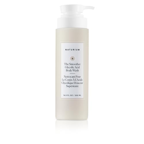
Naturium
The Smoother Glycolic Acid Exfoliating Body Wash
body wash · 500 ml · 27 ingredients
- Fragrance-free
- With lactic acid
- With glycolic acid

Altruist
Moisturising Fluid 0.5% Hyaluronic Acid
body lotion · 50 ml · 23 ingredients
- Fragrance-free
- With urea

CeraVe
Hydrating Cream-to-Foam Cleanser
body lotion · 236 ml · 60 ingredients
- Fragrance-free
- With lactic acid
- With salicylic acid
See the full guide: Best Body Lotions for Keratosis Pilaris →
When to see a professional
See a dermatologist if: you are uncertain whether your bumpy skin is KP or another condition (folliculitis, lichen spinulosus, or pityriasis rubra pilaris may resemble KP); if the redness is severe, spreading, or causing significant distress; if KP affects the face prominently and does not improve with appropriate treatment after 8–12 weeks; or if you notice scarring or hair loss over affected areas (which may indicate the rarer keratosis pilaris atrophicans variants, which require specialist management). A clinical examination is sufficient for diagnosis — no biopsy is typically needed.
Frequently asked questions
Is keratosis pilaris treatment evidence still recommended in 2026?
Yes. The 2025 systematic review published via PMC (Cureus) confirms that topical keratolytics — urea, AHAs (lactic and glycolic acid), and BHAs (salicylic acid) — remain the best-supported first-line treatments for KP. The overall evidence base is characterized by small trials, but consistent real-world clinical use and the known mechanisms of these ingredients support their continued use. No new treatment with superior evidence has emerged to displace them as first-line agents.
Can keratosis pilaris be cured permanently?
No — KP is a chronic genetic condition with no permanent cure. Treatment suppresses the keratin plugging while you use it, but the underlying predisposition remains. Many people notice significant natural improvement in their twenties and thirties, and KP on the face in children often resolves spontaneously. Consistent use of keratolytic moisturizers is the most practical long-term management strategy.
Does diet affect keratosis pilaris?
There is no solid clinical evidence linking specific dietary patterns to KP. The condition's root cause is genetic — a filaggrin mutation affecting follicular keratinization, not a nutritional deficiency. While vitamin A is essential for normal skin keratinization, supplementation does not correct the genetic keratin dysregulation in KP. Focus treatment on topical keratolytics rather than dietary changes.
What percentage of urea is best for keratosis pilaris?
Most over-the-counter KP-targeted body lotions use 10–20% urea. At 10%, urea provides good moisturizing and mild keratolytic activity. At 20%, it is more actively keratolytic and better suited to rougher, more resistant plaques. Concentrations above 20% (up to 40%) are prescription-grade and typically reserved for very thick, resistant areas. Start with 10–12% if new to urea; increase to 20% if needed.
Is it safe to use retinoids on keratosis pilaris?
Yes, with appropriate precautions. OTC adapalene 0.1% or prescription tretinoin can be applied to KP-affected areas on alternate nights. They increase skin sensitivity and can cause dryness and peeling initially — start slowly and always use SPF on treated areas. Retinoids are not appropriate in pregnancy. A dermatologist can guide you on the right strength and pacing if OTC options prove insufficient.
Why does my KP get worse in winter?
Cold air holds less moisture than warm air, and indoor heating further dries the environment. Low ambient humidity reduces the water content of the stratum corneum, making follicular keratin plugs more visible and the surrounding skin rougher. Increasing moisturizer application frequency in winter — especially immediately after bathing — and using a humidifier can reduce the seasonal worsening.
Health information, not medical advice.
Beauty Comparison is an independent editorial publication. This page is researched and sourced to the authorities listed below, but it is general information — not a diagnosis or a substitute for personalised advice from a qualified doctor, dermatologist or pharmacist. If your symptoms are severe, spreading, painful, or not improving, see a healthcare professional.Sources
- 01Keratosis Pilaris: Who Gets and Causes — American Academy of Dermatology (AAD). KP is caused by a buildup of keratin that blocks hair follicles; associated with dry skin and genetic predisposition; worsens in low humidity.
- 02Keratosis Pilaris: Diagnosis and Treatment — American Academy of Dermatology (AAD). First-line treatments include moisturizing creams with urea, lactic acid, glycolic acid, or salicylic acid; topical retinoids used for resistant cases; improvement requires months of consistent use.
- 03The Effectiveness of Topical Keratolytics (AHAs/BHAs/Urea) in Treating Keratosis Pilaris: A Review of the Literature — PMC / Cureus, 2025-12-31. Systematic review confirming AHAs, BHAs, and urea all demonstrate benefit for KP roughness reduction; evidence constrained by small samples and heterogeneous designs; keratolytics remain first-line.
- 04Keratosis Pilaris and Filaggrin Loss-of-Function Mutations in Patients with Atopic Dermatitis — PMC / Acta Dermato-Venereologica, 2022-10-01. Finnish cross-sectional study finding significant association between KP and FLG loss-of-function mutations; supports genetic barrier-dysfunction model for KP.
- 05Keratosis Pilaris — DermNet — DermNet NZ. KP clinical features, prevalence estimates (50–80% adolescents, 40% adults), seasonal worsening, association with ichthyosis vulgaris and atopic dermatitis, variant classification.
- 06Treatment of Disseminated Keratosis Pilaris-associated Itch with Dupilumab — PMC / NIH, 2025-01-01. Case evidence for dupilumab improving KP in patients with atopic dermatitis, supporting downstream barrier-restoration mechanism.