Basal Cell Carcinoma: Signs, Causes & When to See a Derm
The short answer
Basal cell carcinoma is the most common skin cancer.
What Is Basal Cell Carcinoma?
Basal cell carcinoma (BCC) is a malignant tumor arising from the basal cells — the deepest layer of keratinocytes in the epidermis. It is the most common skin cancer and the most common malignancy in the United States and most Western countries. The AAD estimates approximately 3.5 million new BCC cases are diagnosed in the US annually, representing around 80% of all skin cancers worldwide.
Despite being a cancer, BCC has a very different clinical trajectory from many malignancies. It grows slowly, tends to remain localized, and has an extremely low metastasis rate — estimated at under 1% in most published literature. The S2k guideline (2023 update), the most recent comprehensive European consensus on BCC management, cites a metastasis rate of 0.0028%–0.55%. This low rate of spread is why most BCCs, when caught and treated early, are highly curable.
The danger of BCC lies not in spread to distant organs but in local invasion. Left untreated for years, BCC can grow downward and sideways — infiltrating cartilage, bone, muscles, and nerves. Tumors on the nose, eyelids, ears, and temples (the "H-zone" or high-risk zone of the face) are particularly capable of causing significant structural and functional damage.
Who Gets Basal Cell Carcinoma?
BCC can develop in anyone but is substantially more common in people with certain risk profiles.
Epidemiology:
- The S2k guideline (2023) cites an annual incidence of approximately 200 per 100,000 in Central and Northern Europe, with lifetime prevalence exceeding 10%
- Mean age of onset is around 73 in men and 71 in women; it is more common in men
- In the US, the large majority of BCCs affect people of European descent, but BCC does occur in all skin tones
- Once you've had one BCC, your risk of another is substantially elevated — follow-up surveillance is lifetime
Primary Risk Factors:
- Cumulative UV radiation — both chronic sun exposure (outdoor workers, those living in high-UV climates) and episodic intense exposure (sunburns) contribute. UV-B radiation in particular causes the characteristic C→T mutations in the PTCH gene
- Indoor tanning — tanning beds deliver concentrated UV-A and UV-B radiation; their use is a modifiable risk factor
- Fair skin, light hair, light eyes — Fitzpatrick skin types I–II have less melanin to absorb UV radiation, making them most vulnerable
- Age — cumulative UV damage accumulates over a lifetime; BCC is most common in people over 50
- Previous skin cancer — a personal history of any skin cancer significantly increases risk of subsequent BCC
- Chronic immunosuppression — organ transplant recipients and people on long-term immunosuppressive medications face 4–7 times higher risk (S2k guideline 2023)
- Genetic conditions — Gorlin syndrome (basal cell naevus syndrome, caused by inherited PTCH mutation) causes multiple BCCs from a young age; xeroderma pigmentosum (impaired DNA repair) causes extreme UV sensitivity
- Ionizing radiation — prior therapeutic radiation to the skin area increases future BCC risk
- Arsenic exposure — historically from pesticides or contaminated water
The Genetic Mechanism: The Hedgehog Pathway
Understanding why BCC is treated with hedgehog pathway inhibitors in advanced disease requires understanding its molecular basis.
BCC's molecular driver is the hedgehog (Hh) signaling pathway — a developmental pathway critical to embryonic skin growth. In normal adult skin, hedgehog signaling is largely dormant. In BCC, it becomes constitutively active (chronically switched on):
- In approximately 90% of BCCs, UV radiation causes an inactivating mutation in PTCH1 (patched-1), the tumor suppressor gene that normally inhibits hedgehog signaling
- In roughly 10% of BCCs, an activating mutation occurs instead in SMO (smoothened), a downstream effector
- The result is unregulated GLI transcription factor activity, driving abnormal cell proliferation
This pathway-specific mechanism is why vismodegib and sonidegib — SMO inhibitors — can achieve responses in locally advanced and metastatic BCC that cannot be resected or irradiated.
How Basal Cell Carcinoma Looks: Clinical Subtypes
BCC does not have a single appearance. It presents in several distinct subtypes, each with characteristic features. Knowing the subtypes matters because morphoeic (sclerosing) BCC, in particular, is often missed due to its innocuous appearance.
Nodular BCC (Most Common)
The classic presentation, most common on the face — particularly the nose, cheeks, forehead, temples, and around the eyes.
Appearance:
- A pearly, shiny, or translucent bump — often described as having a "pearl-like" quality in reflected light
- Visible surface blood vessels (telangiectasias) that branch across the surface — these dilated capillaries are a strong clinical clue
- May have a central depression or ulcer (the "rodent ulcer" — a historical term for a BCC that has ulcerated centrally)
- Color: typically pink, flesh-colored, or slightly red on fair skin; may be brown or black in people with darker skin tones
- Texture: firm when palpated
- Tendency to bleed easily after minor trauma and then crust over and partially heal, only to bleed again
Superficial BCC
The most common subtype on the trunk and shoulders, particularly in younger patients (30s–40s).
Appearance:
- A flat or slightly raised, scaly, red or pink patch — may look like eczema, psoriasis, or a superficial scar
- Borders often relatively well-defined, sometimes with a slightly raised, thread-like edge
- Less dramatic than nodular BCC; often overlooked or attributed to a benign dermatosis for months or years
Morphoeic (Sclerosing) BCC
The most aggressive and most commonly misdiagnosed subtype. Its innocuous appearance is its danger.
Appearance:
- A pale, waxy, or ivory-colored scar-like plaque with indistinct borders
- Flat or slightly sunken rather than raised; may look like an old scar
- Borders are difficult to define clinically and often extend well beyond what is visible — this histological "iceberg" pattern makes it the subtype most requiring Mohs surgery
- Often diagnosed late because it does not look alarming
Pigmented BCC
A variant of nodular or superficial BCC containing melanin pigment. More common in people with darker skin tones.
Appearance:
- Brown, dark blue, or black coloring — can closely mimic a melanoma or a benign mole
- The underlying BCC structure (pearly quality, telangiectasias) may still be visible under dermoscopy
Where BCC Appears: Anatomical Distribution
The vast majority of BCCs occur on sun-exposed skin:
- Head and neck: approximately 80% of BCCs — the nose (most common single site), forehead, cheeks, eyelids, periorbital area, temples, ears, and scalp
- Trunk: Superficial BCC is especially common on the back and chest
- Upper extremities: Hands and forearms
- Less common: Legs, feet, and genitals; very rare on the palms and soles
The S2k guideline identifies anatomical "risk zones" based on BCC recurrence probability:
- H-zone (high risk): Nose, eyelids, periorbital region, ears, temples, lips, jaw angles, genitals, hands, feet
- M-zone (medium risk): Cheeks, forehead, chin, scalp, neck
- L-zone (lower risk): Trunk and extremities
Diagnosis
Clinical diagnosis relies on visual examination and dermoscopy. The key dermoscopic features of BCC — identified by a trained dermatologist using a dermatoscope — include:
- Arborizing (branching) telangiectasias — the most specific dermoscopic sign of BCC; large, irregular, branching blood vessels
- Blue-gray ovoid nests — round or oval aggregates of pigment below the surface
- Leaf-like areas and spoke-wheel structures — pigmented patterns indicating follicular differentiation (more common in pigmented BCC)
- Shiny white structures under polarized dermoscopy
Histological confirmation is mandatory. The S2k guideline states that "diagnosis of BCC shall be confirmed by histological examination" following biopsy or excision. No clinical or dermoscopic appearance alone suffices to definitively confirm BCC — only tissue histopathology by a dermatopathologist provides the definitive diagnosis and subtype classification needed to guide treatment.
Treatment Options
Treatment selection is guided by BCC subtype, size, anatomical location, patient immune status, and whether the tumor is primary or recurrent.
First-Line: Surgical Excision
Standard surgical excision with clear histological margins is the primary treatment for most BCCs. The S2k guideline (2023) specifies:
- Low-risk BCCs: 3–5mm clinical margins with conventional histological confirmation
- High-risk BCCs (H-zone, large, aggressive histology, recurrent): >5mm margins, or microscopically-controlled surgery
Reported 5-year recurrence rates after excision are 2–8% for primary BCC.
Gold Standard for High-Risk Lesions: Mohs Micrographic Surgery
Mohs micrographic surgery is the standard of care for BCC in the following situations:
- H-zone location — nose, eyelids, ears, temples, lips (where tissue preservation is critical)
- Morphoeic or infiltrative histological subtype (aggressive growth, difficult margins)
- Large tumors or those with indistinct borders
- Recurrent BCC (after prior treatment)
- BCCs in immunosuppressed patients
Mohs surgery removes tissue in stages, with immediate microscopic margin assessment after each layer — allowing the surgeon to map exactly where residual tumor remains and remove only what is necessary. Published cure rates approach 99% for primary BCC, making it the most efficacious individual treatment.
Non-Surgical Options
Non-surgical treatments are primarily indicated for superficial BCC, elderly or medically unfit patients, or cosmetically sensitive locations where excision would cause significant defect:
| Treatment | Best Indication | Notes |
|---|---|---|
| Imiquimod 5% cream (6-week course) | Superficial BCC on trunk/extremities | Clearance rates 43–100% in published data; can cause significant local inflammation |
| 5-Fluorouracil (5-FU) topical | Superficial BCC | Similar efficacy to imiquimod |
| Photodynamic therapy (PDT) | Superficial and thin nodular BCC | Good cosmetic outcomes; requires pain management |
| Cryotherapy | Small, superficial, low-risk BCCs | Less precise than excision |
| Radiation therapy | Unresectable tumors, elderly patients | Clinical control 92–99% for small tumors; secondary malignancy risk after 20 years means it is avoided in younger patients |
| Electrodesiccation and curettage (ED&C) | Low-risk, small lesions on trunk | 93–98% efficacy; produces visible scarring |
Systemic Therapy for Advanced Disease
When BCC cannot be treated with surgery or radiation — locally advanced disease growing into vital structures, or the rare case of metastatic BCC — systemic therapy is required:
Hedgehog pathway inhibitors (first-line):
- Vismodegib (Erivedge): 150mg orally once daily. Achieves 48% response rates in locally advanced BCC and 33% in metastatic BCC (S2k guideline 2023 data). Significant side effects include muscle cramps, alopecia, dysgeusia (altered taste), fatigue, and weight loss. Intermittent dosing protocols (12 weeks on, 8 weeks off) improve tolerability.
- Sonidegib (Odomzo): 200mg orally once daily. Similar mechanism and side-effect profile.
Immunotherapy (second-line):
- Cemiplimab (Libtayo): FDA-approved anti-PD-1 antibody for BCC that has progressed on or is intolerant to hedgehog inhibitor therapy. A 31% objective response rate with 6% complete remission reported in clinical data for this population.
Prevention
The primary driver of BCC is UV radiation, making photoprotection the cornerstone of prevention:
- Daily broad-spectrum SPF 30+ sunscreen — on all exposed skin, every day, regardless of weather. Zinc oxide and titanium dioxide provide the most consistent UV-A and UV-B coverage.
- Protective clothing — UPF-rated shirts and pants, wide-brimmed hats (minimum 3-inch brim to protect ears and neck), UV-blocking sunglasses (wrap-around style protects periorbital skin)
- Avoiding peak UV hours — 10am–4pm when UV index is highest
- No tanning beds — the WHO classifies tanning devices as Group 1 carcinogens; there is no safe level of indoor UV tanning
- Nicotinamide (vitamin B3) supplementation: A randomized controlled trial published in the New England Journal of Medicine (Chen et al., 2015) demonstrated that 500mg nicotinamide twice daily reduced new BCC and squamous cell carcinoma counts by 23% in high-risk patients (those with ≥2 prior non-melanoma skin cancers in the past 5 years). Nicotinamide is not a substitute for sunscreen but is a documented adjunct for high-risk individuals.
Follow-Up: Surveillance After BCC Treatment
After a BCC is successfully treated, ongoing surveillance is essential. The S2k guideline (2023) recommends:
- Low-risk, surgically-treated BCC: Follow-up examination at 6 months after treatment, then annually
- Multiple BCCs, high-risk features, or advanced disease: Every 3 months; annual surveillance after 2 years without recurrence
- Self-examination: Monthly self-checks to detect new BCCs or signs of recurrence at the prior treatment site
Having one BCC meaningfully increases the probability of developing another. This is not a reason for alarm — it is a reason for continued vigilance and regular professional skin checks.
Products that fit: sensitive skin
Picked by code from each product's full ingredient list.

Anua
3 Ceramide Panthenol Moisture Barrier Cream
moisturizer · 100 ml · 63 ingredients
- Fragrance-free
- No essential oils
- No drying alcohol
- No MI/MCI
- No menthol, camphor and witch hazel

Naturium
Marshmallow Root Barrier Balm
moisturizer · 50 g · 58 ingredients
- Fragrance-free
- No essential oils
- No drying alcohol
- No MI/MCI
- No menthol, camphor and witch hazel
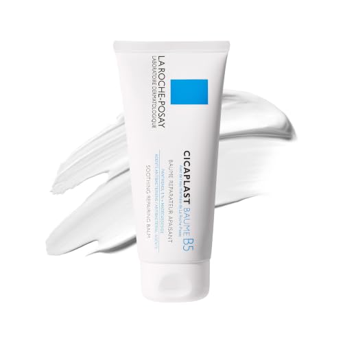
La Roche-Posay
Cicaplast Baume B5 Soothing Repair
moisturizer · 100 ml · 33 ingredients
- Fragrance-free
- No essential oils
- No drying alcohol
- No MI/MCI
- No menthol, camphor and witch hazel

Beauty of Joseon
Red Bean Water Gel
moisturizer · 99.9 ml · 58 ingredients
- Fragrance-free
- No essential oils
- No drying alcohol
- No MI/MCI
- No menthol, camphor and witch hazel
When to see a professional
See a board-certified dermatologist promptly — within days to a week or two — if you notice: a pearly, shiny, or translucent bump on sun-exposed skin; a sore that bleeds easily and fails to heal within 3–4 weeks; a flat, pale, scar-like area you cannot explain; a scaly pink or red patch that doesn't resolve with moisturizer after 4–6 weeks; or any new skin lesion on sun-exposed skin in someone over 40 or with prior skin cancers. Do not adopt a 'wait and see' approach. BCC does not self-resolve and does not stop growing. Early diagnosis and treatment reduces tissue destruction, scarring, and the complexity of the repair procedure needed. Annual professional full-body skin examinations are warranted for everyone with a history of BCC.
Frequently asked questions
Is basal cell carcinoma still highly treatable in 2026?
Yes. When diagnosed early and treated by a dermatologist, BCC has a high cure rate — Mohs micrographic surgery achieves approximately 99% cure rates for primary BCC in high-risk locations. Standard excision achieves 5-year recurrence rates of 2–8%. The 2023 updated S2k guideline — the most comprehensive current consensus on BCC management — confirms surgical excision with margin control as the effective primary treatment, with hedgehog pathway inhibitors (vismodegib, sonidegib) and immunotherapy (cemiplimab) as effective options for the small subset of cases that cannot be surgically treated. The key variable is early diagnosis: BCC treated when small and localized requires only a straightforward office procedure; BCC left to grow for years may require complex reconstruction and still carry risk of recurrence.
Can basal cell carcinoma spread to other organs?
Metastatic BCC is extremely rare — published estimates range from 0.0028% to 0.55% of cases (S2k guideline 2023). When it does occur, it is typically in people with very large, neglected tumors, those with immunosuppression, or those with the basosquamous (mixed) histological subtype, which has more aggressive behavior than pure BCC. The primary concern with untreated BCC is not distant spread but local destruction — BCC can invade cartilage, bone, nerves, and orbital structures if left to grow for years. Rare but documented cases of BCC invading the skull base, orbit, or dura have been reported. This is why early treatment is important even though metastasis is rare.
What does a basal cell carcinoma feel like?
Most patients first notice a BCC because it behaves unusually — it bleeds easily with minor trauma and then appears to heal, only to bleed again with subsequent minor contact. It may crust over repeatedly without fully resolving. Nodular BCC is often firm to the touch; morphoeic BCC may feel like a firm, slightly depressed scar. BCCs are typically painless unless they have grown into or around nerve tissue, in which case patients may notice numbness, tingling, or pain. The bleeding-and-partial-healing cycle is one of the most characteristic clinical features and one of the most useful patient-reported symptoms for prompting evaluation.
What is the difference between BCC and squamous cell carcinoma?
Both arise from keratinocytes (non-pigmented skin cells) and are classified as non-melanoma skin cancers. BCC arises from basal cells (deepest epidermal layer) and is driven primarily by the hedgehog pathway; it almost never metastasizes. SCC arises from squamous cells (middle epidermal layer), has a higher metastatic potential — particularly in immunocompromised patients — and tends to appear rougher, more scaly, and more ulcerated. SCC is also more strongly associated with actinic keratoses (UV-damaged pre-cancerous patches) as precursor lesions, whereas BCC tends to arise de novo. Both require surgical treatment, but SCC carries a higher risk of regional lymph node spread and is monitored more closely.
When should I see a dermatologist about a potential BCC?
See a dermatologist promptly — within a week or two — if you notice: a pearly, shiny, or translucent bump on sun-exposed skin; a sore that bleeds easily and repeatedly without healing; a flat, pale, scar-like area that you cannot account for; a scaly red patch on the trunk or face that doesn't resolve with moisturizer; or any lesion that has been present and growing for more than 4–6 weeks. Do not wait months to see if it resolves. BCC does not self-resolve. The longer it grows before treatment, the more tissue is involved and the more complex the repair. Early BCC is treated with a brief, in-office procedure; advanced BCC may require significant surgery or systemic therapy.
Health information, not medical advice.
Beauty Comparison is an independent editorial publication. This page is researched and sourced to the authorities listed below, but it is general information — not a diagnosis or a substitute for personalised advice from a qualified doctor, dermatologist or pharmacist. If your symptoms are severe, spreading, painful, or not improving, see a healthcare professional.Sources
- 01S2k Guideline Basal Cell Carcinoma of the Skin (Update 2023) — PubMed Central / NIH — Journal der Deutschen Dermatologischen Gesellschaft, 2023-12-01. BCC incidence ~200 per 100,000 annually in Central Europe; metastasis rate 0.0028%–0.55%; 90% of BCCs caused by inactivating PTCH1 mutation; 10% by activating SMO mutation; first-line is complete surgical excision; Mohs surgery for H-zone/high-risk/recurrent BCCs; 3–5mm margins for low-risk; vismodegib achieves 48% response in locally advanced BCC; cemiplimab second-line after hedgehog inhibitor failure; follow-up protocol (6-month then annual for low-risk; 3-monthly for high-risk); immunosuppression increases BCC risk 4–7 fold.
- 02Basal Cell Carcinoma: Symptoms, Causes, and Treatment — DermNet NZ. BCC clinical subtypes include nodular (pearly bump, telangiectasias, central ulceration), superficial (scaly patch, trunk), and morphoeic (waxy scar-like plaque with indistinct borders); dermoscopic signs include arborizing vessels and blue-gray ovoid nests; basosquamous carcinoma is a more aggressive mixed variant; treatment includes excision, Mohs, PDT, imiquimod, and hedgehog inhibitors for advanced disease.
- 03Comprehensive Insights Into Basal Cell Carcinoma: Causes, Presentation, Prevention, and Modern Therapeutic Approaches — PubMed Central / NIH. BCC represents approximately 80% of all skin cancers worldwide; 3.5 million new US cases annually; UV radiation is the primary cause in fair-skinned individuals; Mohs surgery achieves ~99% cure rates; dermoscopy improves pigmented BCC diagnosis in skin of color; nicotinamide 500mg twice daily reduces new BCCs/SCCs by 23% per RCT; cemiplimab is FDA-approved for advanced BCC.
- 04Basal Cell Carcinoma — Signs, Symptoms and Risk Factors — American Academy of Dermatology (AAD). BCC is the most common skin cancer; grows slowly and rarely spreads; presents as a firm round growth, shiny bump, scaly patch, non-healing sore, or scar-like mark; colors vary by skin tone (pink/red in light skin, brown/black in dark skin); may bleed, crust, or cause itching/pain/numbness; primary risk factors include UV exposure (sun and tanning beds), age >50, fair skin, light eyes, previous skin cancer, and weakened immune system; common locations: head and neck.
- 05Nicotinamide for Skin-Cancer Chemoprevention — New England Journal of Medicine (NEJM), 2015-10-22. Randomized controlled trial: nicotinamide 500mg twice daily reduced new non-melanoma skin cancers (BCC and SCC combined) by 23% over 12 months in high-risk patients with ≥2 prior non-melanoma skin cancers in the past 5 years; effects were not sustained after stopping supplementation.